Although there are at least 18 species of Malassezia, the only canine species of current significance is Malassezia pachydermatis. This yeast can colonise many breeds of dogs, although Basset Hounds, West Highland White Terriers, Shih Tzus, English Setters, American Cocker Spaniels, Boxers, Dachshunds, Poodles and Australian Silky Terriers are predisposed (Bond et al., 2018). In addition, atopy is often associated with secondary Malassezia infection and in these dogs, there is often immediate test reactivity to M. pachydermatis.

Apart from breed predisposition and atopy, there are additional predisposing factors:
- Fleabite hypersensitivity (Figure 1)
- Food hypersensitivity
- Primary and secondary cornification defects
- Folds (increased warmth and humidity favours yeast infection)
- Endocrine disease
- Climate (warm humid climates favour yeast infection)
Some cases are idiopathic, however, and in these, control, but not cure, will be necessary.
Clinical presentation in dogs

- Lesions are most often seen in interdigital spaces, ventral neck,
axillae, perineal region, the external ear canal or folds (Hnilica and
Patterson, 2017) - Moderate to severe pruritus
- Erythema (with or without papules)
- Scaling
- Greasy exudation
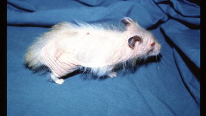
- Hyperpigmentation/lichenification (Figure 2); leathery, elephant-like skin
- Malodour

- Paronychia with crusting, greasy exudation and hyperpigmentation (Figure 3)
- Cheilitis/muzzle erythema; these cases often exhibit extreme facial pruritus
Approach to the case
A full history should be taken, physical examination performed, and clinical signs noted. Cytological examination should be performed. A variable number of yeasts may be present; even small numbers with supporting clinical signs should prompt initiation of therapy, though resampling may be necessary if doubt exists.
The most useful sampling technique in practice is adhesive tape, and the best results are obtained with ultratransparent tape. To do this, apply the tape to the skin, remove it, wrap it around the glass slide for staining, Diff- Quik stain it and then examine it under oil immersion high power to identify yeast organisms (Figure 4).

The number of yeast organisms varies according to the breed, site sampled, technique used and host immune status. More organisms are isolated with tape strips than dry scrapings. Numbers are higher in the axilla region, and lower if hypersensitivity has developed. Bassets do not exhibit hypersensitivity and numbers can be particularly high in this breed. Contact plates and detergent cup scrubs allow quantitative culture – but these are more frequently used in research.
Relying on M. pachydermatis counts for diagnosis is problematical because, although tape stripping is the most widely used diagnostic test in primary care practice, the number of organisms seen is variable and has a low sensitivity. Furthermore, there are, as mentioned, breed and site variations, as well as overlap between counts in health and disease. Thus, a threshold population has not been identified.
Many clinicians employ trial therapy where clinical signs and/or the presence of yeast organisms strongly suggest Malassezia dermatitis. Response to therapy (partial or complete) is usually seen within three weeks.

Histopathological examination
It is debatable whether histopathological examination is more reliable than tape stripping in the primary care setting. Organisms may or may not be seen in the deeper regions of the stratum corneum (Figure 5).
Therapy
Therapy may be systemic, topical or a combination of both.
Systemic therapy
Malassezia dermatitis will respond to treatment with any of the drugs listed below. The best evidence of efficacy is with ketoconazole or itraconazole (which is the most frequently used systemic treatment in the UK currently).
- Ketoconazole (5 to 10mg/kg sid/bid)
- Itraconazole (5 to 10mg/kg sid or two days on, five days off)
- Fluconazole (5 to 10mg/kg sid) plus cefalexin
- Terbinafine (30mg/kg sid or pulse) with or without cefalexin
Topical therapy
Topical therapy is the initial recommended treatment in most cases. Best evidence to date is for 2 percent miconazole plus 2 percent chlorhexidine, applied twice weekly with a contact time of at least 10 minutes. Other treatments reported to be successful are:
- 3 percent chlorhexidine shampoo twice weekly
- Chlorhexidine with or without climbazole mousse (applied every three days) can be alternated with shampoo; this is convenient for owners and there are anecdotal reports of efficacy
Response to treatment may be complete – test for underlying cause, or partial – test for residual disease, eg pyoderma and then underlying cause.
Prevention
Correct the underlying disease and consider intermittent topical therapy; this is the preferred option for long-term therapy. Also consider pulse treatment (but note that this may favour development of resistance) and allergen-specific immunotherapy. Note however that there is limited evidence to date of the efficacy of any of the above.
Zoonotic aspects
There have been several isolated reports of nosocomial infection with M. pachydermatis, especially in neonatal baby units. Overall, the risk appears to be low, provided that barrier nursing and hand hygiene is of a high standard.
Summary
Malassezia should be considered routinely in canine dermatitis. Always do sampling for cytological examination in any case suspected on clinical grounds. Treat yeast when it is found and gauge the effect (clinically and mycologically). Use topical first line where practicable and systemic (itraconazole as first choice) for the “unbathable” or severe cases. Assess concurrent predisposing factors throughout.
| New, extremely comprehensive, open access guidelines are available for Malassezia dermatitis, issued by a panel of WAVD specialists. |


-1628179421-401x226.jpg)