PAPILLOMAS are benign epithelial tumours caused by infection with species-specific DNA papillomaviruses (Hnilica, 2011). The viruses may be transmitted by direct contact or via fomites. The incubation period varies between one and two months.
In the skin there are various types of papilloma.
- Cutaneous papilloma. These are more common in older dogs. Lesions are generally less than 0.5cm in diameter and affect the head, eyelids and feet predominantly. Single or multiple papillomata are possible and they vary in form from pigmented, smooth, alopecic or pedunculated masses. A variant is cutaneous inverted papilloma, usually seen in young dogs on the ventral abdomen and inguinal area as round, raised, single or multiple masses 1-2cm in diameter.
- Multiple pigmented plaques. These occur most commonly in young adult Miniature schnauzers and Pugs. Lesions begin as pigmented plaques and develop into hyperkeratotic masses on the ventrum and medial thighs (Hnilica, 2011). They do not regress and may progress to squamous cell carcinoma.
- Infrequently, papillomata may arise on the penile or vaginal mucosae, and on the footpads.
Canine oral papillomatosis
- Oral papillomatosis occurs in young dogs most commonly.
- Affects the oral cavity and lips.
- Other areas less commonly affected include the nasal planum, eyelids, pharynx, epiglottis and cornea (Miller, Griffin and Campbell, 2013).
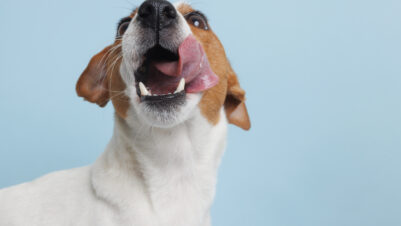
- Lesions begin as smooth papules a few millimetres in diameter and progress to large pedunculated cauliflower-like masses up to 3cm in diameter (Figures 1 and 2).
- Immunosuppression with glucocorticoids or cyclosporine A, or following cancer chemotherapy, may predispose to papillomata or prolong the time to natural remission.
Diagnosis
- The differential diagnosis includes other epithelial neoplasms.
- Physical examination is usually sufficient as the lesions and age of the affected dog are typical.
- Confirmation of the diagnosis may be made, if doubt exists, via histopathological examination, immunohistochemistry or PCR, although these are rarely necessary.
Treatment
The majority of papillomata regress spontaneously. Oral papillomata usually regress within three months (occasionally longer). Cutaneous lesions may persist for 6 to 12 months. Spontaneous remission is as a result of the host cellmediated response.
- Oral lesions may be observed without treatment unless they are irritating the dog, interfering with eating, or are unsightly.
- In these cases treatment comprises surgical removal of the lesions causing problems, with others left alone.
- Ablation using laser or cryotherapy has been used successfully but multiple treatments are usually required (Paterson, 2008).
- Autogenous vaccines and immunomodulating drugs (levamisole, thiabendazole, for example) have been advocated but evidence for their efficacy is lacking.
- A recombinant canine oral papillomavirus vaccine has been produced in the United States and shows promise for the treatment of the rare case of canine oral papillomatosis that is refractory (Hnilica, 2013).
References and suggested reading
Hnilica, K. A. (2013) Papillomas: In: Small Animal Dermatology – A Color atlas and Therapeutic Guide, 3rd ed. Elsevier: pp161-162.
Miller, W. H., Griffin, C. G. and Campbell, K. L. (2013) Papilloma: In: Muller and Kirk’s Small Animal Dermatology, 7th ed. Elsevier: pp774- 778.
Paterson, S. (2008) Papillomavirus: In: Manual of Skin Diseases of the Dog and Cat, 2nd ed. Blackwell Publishing: pp84-86.