Prolapse of tissue from the cloaca is a true emergency in reptiles. The cloaca has variable anatomy between reptile species; however, broadly it is an external opening for the gastrointestinal, urinary and reproductive tracts. Any prolapse of tissue from the cloaca will involve one or more of these systems. Prolapse of mucosal tissue from the cloaca will result in rapid mucosal devitalisation and ischaemic necrosis (McArthur and Machin, 2019a). If prolapsed tissue is not protected and reperfused swiftly, necrosis of the tissue and, potentially, death of the patient can result. Cloacal prolapse has been reported in numerous reptile species but is reported as more frequent in chelonians and chameleons (McArthur et al., 2004). A retrospective study of reptile cloacal prolapses over 14 years showed chelonians and lizards were 3.47 times more likely to present with a cloacal prolapse compared to snakes (Hedley and Eatwell, 2014).
A retrospective study of reptile cloacal prolapses over 14 years showed chelonians and lizards were 3.47 times more likely to present with a cloacal prolapse compared to snakes
Initial examination
With the exception of intermittent protrusion of the phallus or clitoris through the cloaca in chelonians, any protruding tissue should be assessed in the veterinary clinic as soon as possible (McArthur and Machin, 2019a). The client should be instructed to moisten the protruding tissue with a water-based lubricant before making their way swiftly to the veterinary practice.
On presentation, a full assessment of the prolapsed tissue and the patient’s overall health as well as a full husbandry review should be performed. It is essential to remember that cloacal prolapses are a symptom rather than a primary problem, and are the result of underlying disease or husbandry problems. It is also important to identify the prolapsed tissue, if possible. This may not always be immediately achievable, as tissue that has been prolapsed for a prolonged period of time can be severely oedematous, devitalised or deformed (Figure 1); additionally, the tissue may have been traumatised by the patient.
Identification of the prolapsed tissue can be difficult; however, assessing for the presence of a lumen can help to differentiate colon or oviduct from hemipene, phallus, bladder or a prolapsed mass
Potential prolapsed structures include cloacal tissue, colon, intestine, bladder (in those species with a bladder) (Figure 2), oviduct, phallus or hemipenes (McArthur and Machin, 2019a). Identification of the prolapsed tissue can be difficult; however, assessing for the presence of a lumen can help to differentiate colon or oviduct from hemipene, phallus, bladder or a prolapsed mass. Longitudinal striations can help to differentiate oviduct from other organs (Figure 3), as these are not present on cloacal or colonic tissue (McArthur and Machin, 2019a). In one study, the most commonly reported prolapsed tissue was a phallus or hemipene, representing 35.7 percent of all cases (Hedley and Eatwell, 2014).


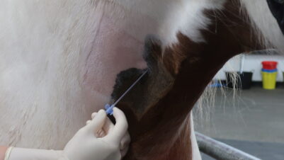
During examination, patients should be provided with analgesia, as cloacal prolapses are painful and distressing (McArthur and Machin, 2019b). Potential options for analgesia include systemic opioids, tramadol or NSAIDs, ideally in combination with local anaesthesia (McArthur and Machin, 2019a). If possible, an epidural can be considered to help minimise straining and relax the cloacal structures to aid replacement of the prolapse.
If not already performed by the client, the prolapsed tissue should be lubricated, and gentle cleaning with warm water or saline should be commenced if any gross contamination is present. Assessment of the prolapsed tissue should ideally include determining the type of tissue or organ of origin, whether the tissue is traumatised or irreversibly desiccated and whether replacement of the tissue is feasible or if it requires amputation. In the case of phallus or hemipene prolapse this is relatively straightforward. However, amputation of cloacal or oviductal tissue is difficult and requires coeliotomy, while amputation of a prolapsed bladder should not be attempted.
Diagnosis
Cloacal prolapses can be caused by any number of underlying issues, often associated with excessive straining. More specifically, common causes of cloacal prolapse include dystocia, gastrointestinal parasitism, urolithiasis, constipation or faecoliths, trauma, chronic sexual activity, coelomic masses, coelomitis or ascites (McArthur and Machin, 2019a). Diagnostic work-up should always be performed on patients with cloacal prolapses, as if the underlying cause is not identified and treated, the prolapse is likely to reoccur. If possible, diagnostic work-up can be delayed until after a prolapse is reduced and secured in place with cloacal retention sutures.
As the cloaca is a slit-like opening, purse-string sutures cannot be placed as for a canine or feline patient, so sutures must be placed on the lateral aspect of the cloacal opening bilaterally
Use of hypertonic saline or cold compresses can aid in reducing the size of viable prolapsed tissue so that it may be replaced (Sykes, 2010). As the cloaca is a slit-like opening, purse-string sutures cannot be placed as for a canine or feline patient, so sutures must be placed on the lateral aspect of the cloacal opening bilaterally. Care should be taken when manipulating prolapsed tissue to be as gentle as possible, as it is often friable (Music and Strunk, 2016).
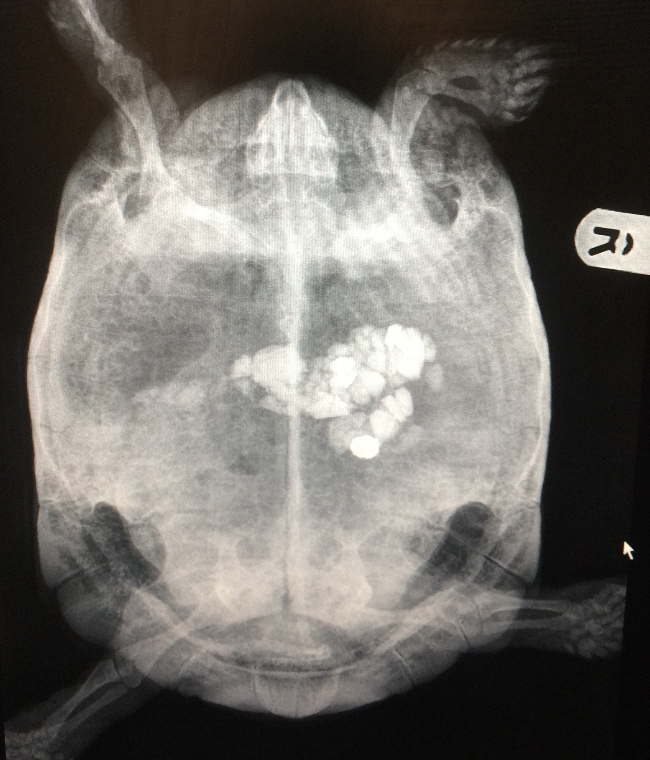
If replacement of the prolapsed tissue is not possible at first presentation, the use of protective wraps such as cling film or plastic wrap can help keep hypertonic saline soaked swabs or lubricant in place, preventing trauma to the prolapsed tissue (McArthur and Machin, 2019a). Initial work-up should always include imaging (McArthur and Machin, 2019a). Radiographs can help identify metabolic bone disease, urolithiasis, presence of eggs (Figure 4) and severe constipation (Figure 5). Other diagnostics to consider include faecal parasitology, biochemistry and haematology, and ultrasound examination (McArthur and Machin, 2019b).

Treatment and prognosis
As management of cloacal prolapses often involves concurrent medical treatment for the underlying condition and surgical treatment for the prolapse, it is important to ensure the client is fully on board with understanding and treating the underlying cause as well as the presenting condition
The prolapsed organ(s) often determine the treatment and prognosis of the condition. As management of cloacal prolapses often involves concurrent medical treatment for the underlying condition and surgical treatment for the prolapse, it is important to ensure the client is fully on board with understanding and treating the underlying cause as well as the presenting condition (McArthur and Machin, 2019b). Prognosis depends on numerous factors, including the organ that is prolapsed, the viability of the tissue, the underlying cause and the overall health of the patient (McArthur and Machin, 2019a).
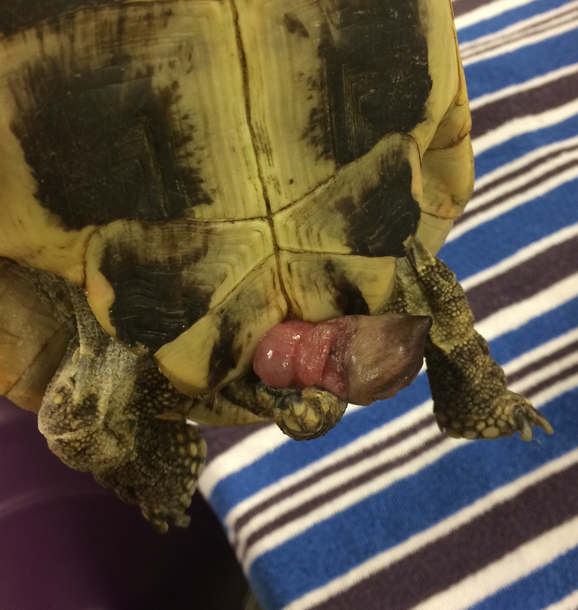
Generally, hemipene or phallus prolapses (Figure 6) have an excellent prognosis, as they only play a role in sexual reproduction and can be amputated in a brief surgical procedure. The patient should be sedated or anaesthetised and, if possible, the phallus or hemipene should be replaced and retention sutures applied. If the tissue is too devitalised or damaged, the phallus can be clamped at the base, ligated with encircling ligatures and amputated under anaesthesia (Sykes, 2010). The author prefers to perform this procedure with alfaxalone induction and the use of local anaesthesia.


Prolapse of other organs is often less straightforward and carries a poorer prognosis. Coeliotomy is often required to correct prolapse of colon, oviduct or bladder (McArthur and Machin, 2019b). If the tissue is devitalised, amputation is often required. This can involve resection and anastomosis of cloacal or colonic tissue, or removal of the entire oviduct. Most patients with oviductal, colonic or bladder prolapse fail to return to a normal quality of life (McArthur and Machin, 2019a). In cases of more complicated prolapse, veterinarians should consider referral for further evaluation or euthanasia.
Summary
Cloacal prolapse is a clinical sign of underlying illness and should be treated as a symptom rather than a disease entity. It is essential to determine the underlying cause to prevent re-prolapse of the tissue. Initial assessment should determine the organ that has prolapsed and the viability of the tissue. Prognosis depends on many factors, including the underlying health of the patient and assessment of the prolapse, as well as the underlying cause.