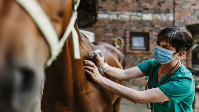
Although most colic episodes in horses resolve with or without basic medical therapy, approximately 7 percent will require surgical correction (Archer, 2004). One of the most important questions facing a first opinion veterinarian examining a horse with colic in the field is whether referral to a veterinary hospital with surgical facilities is required. It may be that referral is not an option for the owner, in which case the decision-making process is simplified. If referral is an option, the attending veterinarian will need to assess the patient, looking for signs that a lesion requires more intensive treatment or surgery. This article will discuss this decision-making process (Figure 1).

Important considerations for referral
It is important to be aware of the British Equine Veterinary Association guidelines for euthanasia where horses are insured for mortality. In cases where a horse requires urgent surgical or specialised medical intervention to save its life, it is important to keep the insurance company notified wherever possible, although it is recognised this may not always be practical out of normal business hours. There will also be circumstances, for example, where the estimated cost of treatment may not be economical when compared to the horse’s value. These are matters of negotiation between the insurance company and owner and are not for the veterinarian to decide.
Costs
The costs of referral should be relayed to the client. At the time of writing this article, a medically managed colic would typically cost £1,500 to £4,000, while a surgically managed colic would typically cost £4,000 to £7,000.
Initial examination
There are numerous causes of colic, most of which are related to the gastrointestinal tract. Non-intestinal causes of colic and colic-like symptoms include:
- issues with the reproductive system such as metritis, uterine artery haemorrhage and uterine torsion
- musculoskeletal pain (rhabdomyolysis and laminitis)
- neurological disease
- hepatic disease
- urinary tract disorders
- pleuritis
- cardiac conditions
A targeted history and thorough clinical examination will help rule these in or out.
The initial exam will necessarily be brief as you will likely be dealing with a horse in pain with pressure from the owner to treat them quickly. A history can be taken while the horse is being examined. The duration of signs (when the horse was last seen normal) and their intensity (worsening or the same), any response to drugs administered (the owner may have administered oral phenylbutazone or similar), faecal output, worming history, recent changes of management and whether the horse is pregnant should all be ascertained. An observation of the degree of pain, measurement of heart rate and auscultation for the presence of gut sounds should ideally be assessed before the administration of pain relief, which may affect these factors.
Once these parameters have been collected, a full dose of a non-steroidal anti-inflammatory drug (NSAID) should be given intravenously. Opinions differ regarding the choice of NSAID, but in the author’s opinion, it has little bearing on the decision-making process for colic referrals. Concurrent administration of an antispasmodic (N-butylscopolammonium bromide at 0.1mg/kg IV) also facilitates subsequent rectal examination and improves safety. If the horse is particularly painful, concurrent administration of an alpha-2 agonist with or without butorphanol will provide more rapid visceral pain relief. However, the author prefers xylazine (100 to 200mg in a 500kg horse).
Gastric distension is very painful, and relief of the pressure will have a significant effect on the degree of pain
If the heart rate prior to sedation is elevated (over 52 bpm), a nasogastric tube should be passed to check for the presence of reflux. This is of vital importance as horses cannot vomit, so fluid and gas can accumulate in the stomach leading to its rupture. Gastric distension is very painful, and relief of the pressure will have a significant effect on the degree of pain. If more than 2 litres of reflux are obtained, referral should be recommended. The nasogastric tube can be left in place and taped to the headcollar if the horse will need to travel a long distance. An elevated heart rate over 60 bpm should be viewed with high suspicion, and referral should be considered.
Pain assessment
The degree of pain a horse is exhibiting varies and may not accurately reflect the underlying pathology; however, more severe pain is usually associated with severe lesions. Pawing, rolling and sweating are common signs, but some horses suffering from colic will simply stand quite still despite severe pathology. The response to pain relief is important, and failure to respond to a full dose of NSAIDs should prompt referral. Rather than administering further NSAIDs, long-acting alpha-2 agonists (detomidine or romifidine) given intravenously and intramuscularly can allow safe transport of a painful horse.
Diagnostic tests for colic
Rectal examination
As stated earlier, a rectal examination is best performed after administration of an antispasmodic and sedative, if given. There are inherent risks to both the examiner/handler and horse, so safety concerns must be borne in mind. Only 20 to 30 percent of the abdomen can be explored during rectal examination, so not performing the examination in a fractious horse is of little diagnostic consequence. Nevertheless, abnormal findings can be palpated, which assists with the decision on whether to refer.
The small intestine is not usually identifiable, but when distended, it will feel like loops of bike tyre inner tube. If this is palpated, a nasogastric tube should be passed. Large colon distension will be obvious, and a large viscus, which may even prevent advancement of the hand further then the wrist, can sometimes be felt. Such a finding would usually prompt referral.
Caecal impactions can be difficult to treat, and referral should be considered to allow close monitoring and aggressive treatment
Large colon impactions, such as a pelvic flexure impaction, can be palpated as a large “doughy” structure of varying size, and large colon taenial bands course horizontally. Impactions can usually be treated successfully in the field with enteral fluid therapy, although stubborn impactions can require hospitalisation for administration of intravenous fluids and more aggressive enteral fluid therapy. The caecum is on the right side of the abdomen, with vertically aligned taenial bands. Caecal impactions can be difficult to treat, and referral should be considered to allow close monitoring and aggressive treatment.
Abdominocentesis
Abdominocentesis is a useful diagnostic technique which gives a sensitive indication of gut health. It would usually be performed in a referral hospital as the sterility required and risk to the operator increases its risk in the field. Normal peritoneal fluid is clear/light yellow with a total nucleated cell count (TNCC) of less than 5.0 x 10⁹/l and a total protein of less than 25g/l. The presence of a strangulating small intestinal lesion causes the fluid to become darker, the TNCC to increase to greater than 5.0 x 10⁹/l and the total protein to increase to greater than 25g/l. Values in this range should prompt referral. If large colon distension is palpated per rectum, abdominocentesis should not be performed.
Ultrasonography

Ultrasonography can be performed relatively easily in the field on horses suffering from colic and is particularly sensitive for detecting a distended small intestine (Figure 2). Indeed, a fast localised abdominal sonography of horses (FLASH) protocol has been described (Busoni et al., 2011). In a simple technique, the inguinal region is soaked in alcohol, and a convex probe (2.5 to 5MHz) is used to detect the presence of circular loops of intestine with a wall thickness greater than 0.4cm and loop diameter greater than 3cm. Such findings should prompt referral. Likewise, the large intestine wall thickness can be evaluated, with a wall thickness greater than 1cm suggesting oedema.
Blood tests
A simple blood test to evaluate cardiovascular status can be valuable in assessing hydration and evidence of shock in horses with colic. The packed cell volume (PCV) and total protein (TP) will rise in the presence of small intestinal ileus. Plasma lactate measurement has increased in prevalence with the advent of handheld “stable-side” machines (Nieto et al., 2015). Elevation of lactate (greater than 2mmol/l) indicates poor perfusion of tissues and cells working anaerobically (Donawick et al., 1975). Elevations in lactate are also seen in the presence of large intestinal strangulating lesions and are correlated with survival (Johnston et al., 2007).
Conclusion
The slightest suspicion of a strangulating lesion should prompt referral as early surgical correction may prevent the need for resection
The slightest suspicion of a strangulating lesion should prompt referral as early surgical correction may prevent the need for resection. If you’re unsure and the owner is willing, then referral should be recommended. Referral hospitals will always support the decision to refer and will be happy to advise. If referral is not an option, then pain relief should be maintained alongside appropriate supportive and therapeutic care. If the welfare of the horse is being compromised, euthanasia should be performed.
| What to consider when making the decision to refer a horse with colic – Response to pain relief (NSAIDs) – Distended small intestine – More than 2 litres reflux – Heart rate persistently over 60 beats per minute – Recurrent colic – Alterations in peritoneal fluid – Progressive abdominal distension – Progressive deterioration in mucous membrane colour |


