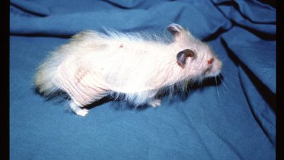
Cheyletiellosis is a skin disease caused by infestation with Cheyletiella mites. There are five species, which are predominantly host specific, although cross infestation may occur:
Rabbits: Cheyletiella parasitovorax (Mégnin, 1878) and Cheyletiella firmani (Smiley, 1970)
Cats: Cheyletiella blakei (Smiley, 1970)
Dogs: Cheyletiella yasguri (Smiley, 1965)
Hares: Cheyletiella strandtmanni (Smiley, 1970)
There are variations in the shape of the solenidion, a specialised chemosensory seta located on the fourth segment of the first leg. The shapes can vary, however, and therefore the precise identification may be difficult, although not of any practical significance as host specificity is thought to be high. C. parasitovorax, C. blakei and C. yasguri are considered the most commonly seen in veterinary practice. The mites spend most of the time in hairs and go to the skin surface to feed on epithelial debris.
The life cycle is of approximately three weeks. Female mites lay eggs that are attached to hairs. Hatching produces larvae followed by the first nymphal stage, a second nymphal stage and then adults. The entire life cycle is spent on the host.
Clinical signs
Clinical signs vary according to the number of mites on the animal and whether hypersensitivity to the bites occurs. The most common sign in non-hypersensitive animals is excess scale. In severe infestations, movements of the mites or associated debris may be seen, hence the term “walking dandruff” (Figure 1). With fewer mites, the scale will be less noticeable. In rabbits the excess scale is dorsal, particularly between the scapulae. In dogs and cats scale may be more generalised, but still predominantly dorsal (Figures 1 and 2).


Pruritus is usually minimal. In animals that develop hypersensitivity there may be severe pruritus and self-inflicted lesions, such as miliary dermatitis or symmetrical alopecia in cats (Figure 3), or mimicking fleabite hypersensitivity in dogs. Due to the current high prevalence of insecticide use in older animals, cheyletiellosis is most likely to be seen in puppies and kittens kept in poor husbandry conditions.

Making a diagnosis
Consider the differential diagnosis of scaling and hypersensitivity. Seborrhoea, poor nutrition, inadequate grooming, demodicosis, pediculosis, otodectic mange and flea infestation may be causes of scaling. In cats, diabetes mellitus, liver disease and other causes of miliary dermatitis may also be considered.
Hypersensitivity can be caused by atopic dermatitis, fleabite hypersensitivity, food hypersensitivity, scabies and ectopic Otodectes cynotis hypersensitivity.
A full history should be taken and a physical examination performed. There may have been no recent antiparasitic treatment or poor compliance. Imidacloprid is an effective antiparasitic agent but is not effective against Cheyletiella. Excess scale is usually obvious and if found, should prompt skin sampling.

Coat brushings should be done, and the debris placed on brown paper or a petri dish. The debris should then be examined using a microscope with a total of X40 magnification. Walking dandruff may be seen, but not necessarily in milder infestations. Acetate tape strips should be taken. Hair should be trimmed and a superficial skin scraping taken and mounted in liquid paraffin.
The adult Cheyletiella, 0.5mm in size, is just visible to the naked eye but microscopic identification is necessary. All legs protrude from the body and end in combs. The mite also has a waist. The most important diagnostic feature is the characteristic hooks of the accessory mouth parts (Figure 4).
Hair plucks can be taken. Cheyletiella eggs will be attached to hairs. In hypersensitive animals it is easier to find eggs than adults. These eggs are attached along the length of the hairs in a fine cocoon. They are smaller than louse eggs and are non-operculate (Figures 5 and 6). Pruritus will cause excessive grooming, especially in cats. In these cases, eggs and adult mites may be detected by routine faecal flotation techniques.

A therapeutic trial, using an acaricidal product administered by a veterinary surgeon or nurse, may be necessary in cases where clinical signs are strongly suspected but the mite or eggs are not found.
The mite will bite in-contact humans causing hypersensitivity wheals, often diagnosed by the family physician as fleabites. These occur in contact sites – arms, waist and legs, for example. Individual members of the owner’s family will not usually develop lesions simultaneously, which may cause confusion. Human lesions depend on the level of contact and whether a hypersensitivity response develops.

Treatment
All in-contact animals should be treated. Cheyletiella is susceptible to many pesticides routinely used for tick and flea control in small animal practice.
Topical products include:
- Lime sulphur dips applied weekly for four weeks
- Fipronil spray (not in rabbits)
- Selenium sulphide shampoo once a week for three weeks
Most spot-on products are also effective, except for imidacloprid. Examples include:
- Ivermectin (Xeno, Dechra) for rabbits
- Selamectin once a month for three months for cats, dogs and rabbits
- Fipronil (dogs and cats only)
- Moxidectin

Oral products used in flea and tick control, such as the recently introduced isoxazolines, or milbemycin are likely to be effective, although as with the other products mentioned, there is no specific licence for Cheyletiella.
Environmental treatment is not strictly necessary, especially if long-acting antiparasitic drugs are used. However, routine cleaning of the environment allied with products designed for flea control is often advised because survival of female mites off the host for up to 10 days has been documented.


-1628179421-401x226.jpg)


