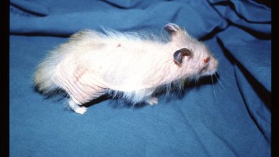
Skin cases benefit from a detailed history, a physical examination to determine the health status of the patient and a dermatological examination. From this, a differential diagnosis list can be formulated, which, once ranked in order of most likely to least likely, can suggest the diagnostic tests necessary to confirm a diagnosis. Ranking is important since it enables tests to be targeted, avoiding the unnecessary expense of tests that may not be relevant to the case in question.
Involvement of veterinary nurses trained in the investigation of skin diseases can be very important. Not only can the nurse be present in the initial consultation to establish a relationship with the owner, and thereby act as a contact for subsequent queries and problem solving, they can also be responsible for the rapid in-house tests necessary for many cases, including the six procedures described in this article.
The equipment required for the six essential tests is minimal, consisting of Diff-Quik stains, glass slides/coverslips, a microscope with low, medium and high power (including oil immersion) and a good quality Wood’s lamp.
Coat brushing

This very simple test, perhaps underutilised in practice, is a useful screen for ectoparasites, eggs, flea farces and follicular abnormalities.
The animal should be brushed over a black consulting table or over
brown paper. The hairs and epithelial debris should be collected and the
hair separated from scale. The hairs are placed in a petri dish
enabling them to be examined with the Wood’s lamp and mounted in liquid
paraffin with a cover slip for microscopic examination. The scale is
also mounted in liquid paraffin and a cover slip applied.
Lice, nits, Cheyletiella and fragmented flea faeces, perhaps missed macroscopically in cases of fleabite hypersensitivity, may be found (Figure 1).
Hair plucks (trichoscopy)
Hair plucks are an inexpensive and rapid test for demodicosis,
especially in areas such as the feet, where skin scraping is difficult.
Hairs are selected from diseased areas determined by the dermatological
examination. Hair at the periphery of lesions is selected, plucked using
tweezers, placed in liquid paraffin and a cover slip applied.


Under low power magnification (x40), Demodex mites (Figure 2) are often found in association with the hair shafts, and the need for more invasive skin scrapings avoided. Cheyletiella may also be diagnosed by hair plucks (either the mite itself, or more frequently, the cocooned eggs) (Figure 3). Apart from parasites, evidence of hair damage from diseases such as dermatophytosis (Figure 4), follicular casts (Figure 2), and evidence of keratinisation problems may be found. In addition, feline pruritus is suggested by broken hair shafts (Figure 5).
Skin scraping
Skin scraping is slightly more time consuming than the previous tests but is essential in cases suspected of parasitic involvement if less invasive tests are negative. It is used primarily to identify surface and burrowing mites causing demodicosis or scabies or, in some cases, other mites such as Cheyletiella.
For demodicosis, deep scraping is required. Clip to remove hair if necessary. Squeeze the skin in the area to be scraped and apply liquid paraffin to the skin. This has the effect of lubricating and penetrating the skin and facilitating collection of material.
Use a blunt blade scrape in the direction of hair growth. The first few scrapes can be superficial and then subsequently deep (enough to cause capillary ooze). Material should be transferred to a slide, mixed with liquid paraffin, a cover slip applied and examination performed under low power.
For scabies, multiple scrapings, both superficial and deep, in non-excoriated areas are required. This is because many cases are associated with hypersensitivity to Sarcoptes scabiei and few mites may be present. Scanning of the slide under low power and not high facilitates the identification of a limited number of mites. Liquid paraffin does not kill the mite and subtle movement may help to locate its position.
Tape strips
Tape stripping is an extremely useful and versatile diagnostic test. It is quick, inexpensive and often yields valuable diagnostic information. It is a technique employed by veterinary dermatologists in the majority of cases, as it can be used to diagnose superficial pyoderma (Figure 6), bacterial overgrowth, some autoimmune diseases and parasites. Tape stripping is the most common technique for diagnosing Malassezia dermatitis or overgrowth (Figure 9).
Several 10cm pieces of tape are pressed onto the surface of the skin.
One sample is examined under low power for parasites such as Demodex, Sarcoptes, Cheyletiella and lice. For the identification of bacteria or Malassezia,
tape is partially attached to the slide and stained with Diff-Quik and
then completely attached for examination under high power with oil
immersion.


Some brands of tape disintegrate with fixative and in these cases, fixation can be dispensed with. Ultra transparent tapes can be fixed without loss of the tape and generally deliver better results.
Slide impressions and swab smears
Slide impressions of the surface of a lesion should be used for any purulent or exudative lesion. This technique is valuable for differentiating between bacterial infection and sterile lesions such as those seen in pemphigus foliaceus (Figures 7 and 8).
Suitable areas for microscopic examination are located under low power and identification of bacteria and Malassezia (Figure 9) is facilitated under high power with oil immersion. The glass slide is pressed against the moist lesion, fixed and stained with Diff-Quik. Alternatively, for purulent material, including pricked pustules, a swab is used to collect material, which is then gently rolled over the surface of the slide.
Wood’s lamp examination
Wood’s lamp examination is a very useful inexpensive screening test for dermatophytosis caused by Microsporum canis only (other dermatophytes of veterinary significance do not fluoresce). It is important that the Wood’s lamp is of a suitable standard from a recognised veterinary supplier. It should produce ultraviolet light at a wavelength of 253.7 nanometres. Good quality Wood’s lamps have two bars and an inbuilt magnifying lens.
Examination must take place in a very dark room. Warm the lamp for five minutes. Scan the animal for a few minutes as sometimes fluorescence is delayed. Position the lamp over hairs collected by brushing, as many dermatophytosis hairs are damaged and therefore easily removed by brushing.

Only infected hairs fluoresce (not surrounding skin or scale) and the
apple green colour is very important as it distinguishes true
fluorescence from false (Figure 10).
Positive fluorescing
hairs can be plucked, examined microscopically and cultured for
confirmation. It is also useful to demonstrate positive fluorescence to
all clinical staff, vets and nurses, as the fluorescence is striking and
educational.
The often-quoted figure that only 50 percent of
M. canis hairs fluoresce is incorrect. Many authorities have achieved
positive results between 80 and 90 percent, with attention to detail
outlined above, although a negative result should not be considered
conclusive.
Thanks to Ross Bond, Anette Loeffler and colleagues from the Royal Veterinary College dermatology service for the use of illustrations.


-1628179421-401x226.jpg)


