


Ectoparasites can be an important cause of disease and welfare concerns in farmed ruminants in the UK and are more common in the autumn and winter. In this article, the conditions that are more likely to be brought to the practising veterinary surgeon’s attention are summarised and other parasitic skin diseases that may be diagnosed in the future are discussed.
Sheep scab (Psoroptes ovis)
This disease is endemic in the UK and notifiable in Scotland and Northern Ireland. Although its prevalence is not known with certainty, it is likely to have increased since the 1990s (Bisdorff et al., 2006). Disease is caused by an allergic reaction (Figures 1 and 2) to Psoroptes ovis, a mite which is capable of infecting sheep and surviving in the environment for approximately 15 days. Sheep scab does not affect humans and rarely affects cattle.


Diagnosis is made by examining skin scrapes and scab material taken from the edge of the lesion. The APHA will examine the sample by direct microscopy (Figure 3), but will also carry out a potassium hydroxide (KOH) digest, which may detect mites if numbers are small, or they are hidden in scab material.
There is also a recently introduced commercial ELISA test that detects antibodies to P. ovis mites. It is to be used in groups of animals and can aid early diagnosis as the antibody response can be detected within two weeks of infection. It can also indicate infection in chronically affected animals where clinical signs and mite numbers may be reduced.
There are only two types of treatment for sheep scab: the organophosphate (OP) dips or the injectable macrocyclic lactones (MLs). All of the affected group should be treated. The ML treatments vary as to their persistency, leading to variations in whether animals have to be moved from the infected areas after treatment (necessary for all ML treatments apart from moxidectin) and the amount of time before they can mix with untreated sheep. They are also all anthelmintics, so their use will have implications for the development of anthelmintic resistance.
The recent publication of detection ofP. ovis mites resistant to MLs on four farms in England and Wales (Doherty et al., 2018) highlights the importance of diagnosis of the cause of pruritus and correct treatment. The Sheep Veterinary Society has published updated guidance on this disease here (opens PDF).
Chewing lice in sheep (Bovicola ovis)
Chewing lice are an important differential diagnosis for sheep scab in pruritic sheep. It is, however, possible to have sheep infested with both Psoroptes ovis mites and Bovicola ovis lice. Diagnosis is by direct microscopic examination of skin scrapes or wool plucks from affected areas.
Treatment of chewing lice in sheep is by OP dip or by topical synthetic pyrethroids. Injectable MLs are not licensed against chewing lice. Topical pyrethroids will only have limited efficacy against lice in full-fleeced sheep.
For further Information, see scops.org.uk/external-parasites/

Psoroptic mange in cattle
Psoroptic mange is an important disease in cattle present in Europe, and outbreaks in the UK (Figure 4) have been linked to imported live animals and bought-in cattle. It is not a notifiable disease in the UK and it does not affect humans. The Psoroptes species found in cattle, though indistinguishable from P. ovis in sheep, appears to be a cattle-adapted strain and has failed to establish in sheep (Jones et al., 2008).
Like the disease in sheep, clinical signs are more apparent in the autumn and winter, allowing sub-clinically infected cattle to be moved at other times. Pruritus and secondary infection (Figure 5) can be severe, particularly in animals receiving poor nutrition and those suffering concurrent disease (Mitchell, 2012).

Diagnosis is by microscopic examination of skin scrapes for Psoroptes spp. mites. Treatment is difficult. Ivermectin injection and moxidectin and doramectin pour-on and injections (all MLs) are licensed for the treatment of psoroptic mange in cattle in the UK, but nothing is licensed for lactating dairy cattle.
However, in the outbreaks investigated in the UK (Jones et al., 2008), the MLs were not fully effective and repeated treatments or alternative products (permethrin pour-on or amitraz) have to be used under veterinary supervision, under the cascade. The treatment should be given to all animals in the group and any in-contact animals. Movement from infected housing should also be advised.
It is very important to repeat the skin scrapes to check the efficacy of the treatment used as clinical signs may improve, but live mites may still be present.
Sucking lice in cattle
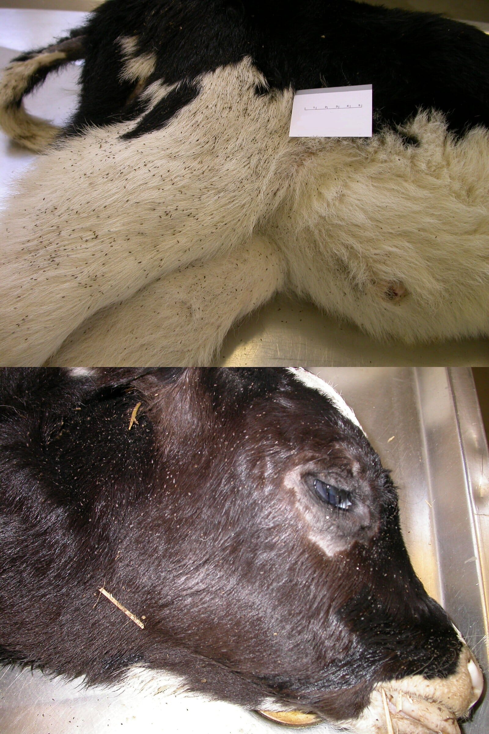
Cattle sucking lice (Linognathus vituli, Solenopotes capillatus or Haematopinus eurysternus) are commonly seen on the head, neck and around the eyes. In young calves, severe infections can cause severe anaemia and malaise and in extreme cases, are associated with death (Figure 6). It is important to remember that a heavy louse infestation may itself be a sign of another underlying condition, such as malnutrition or chronic disease, as debilitated animals may not groom themselves effectively.
Transfer of lice between animals or flocks/herds is usually by direct physical contact as, unlike mites, they cannot survive for long in the environment. Diagnosis can be achieved through microscopic examination to identify adult louse populations and eggs adherent to hairs. Treatment is by pour-on synthetic pyrethroid preparations or injectable or pour-on MLs. All cattle in direct contact must be treated.
For further information, see cattleparasites.org.uk/app/uploads/2018/04/COWS_cattle_parasite_control_guide.pdf
Exotic parasitic skin diseases in cattle
Parafilaria bovicola
Summer “bleeding disease” is caused by a nematode in the subcutaneous tissues of cattle. The nematode punctures through the skin to lay eggs on the skin surface, causing cutaneous bleeding points. Eggs and first-stage larvae are present in the exudate and flies (Musca autumnalis) are infected by feeding. The parasite develops to the infective stage (L3) in the fly and cattle are infected by them feeding on wounds or lachrymal secretions, transmitting the nematode. The disease is present in many European countries.
Besnoitia besnoiti
Besnoitia besnoiti is an emerging protozoan parasite in cattle, present in Europe. It can be spread by biting insects. A proportion of animals show severe clinical signs of progressive thickening, folding or wrinkling of the skin with hair loss. They will also have scleral, conjunctival or vulval cysts.
For further information, see apha.defra.gov.uk/documents/surveillance/diseases/vetinfonote-bovine-besnoitiosis.pdf


