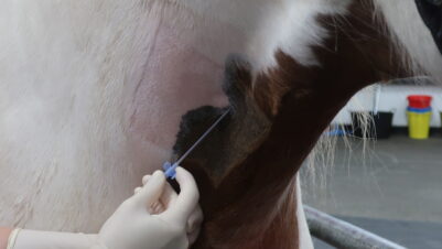
Colic is a serious disorder commonly seen in equine practice. Along with the correct diagnosis and treatment, colic patients require specialist nursing care for the best chance of recovery. However, the registered veterinary nurse (RVN) caring for colic patients must have a good knowledge of the presenting condition, equine behaviour and gastrointestinal (GI) tract anatomy.
Colic
The term colic refers to abdominal pain and is not a specific diagnosis. Table 1 displays the various causes of colic in the horse. Initial assessment aims to separate alimentary causes from non-alimentary causes (Slater and Knowles, 2012).
| Alimentary tract causes | Non-alimentary tract causes | Conditions that resemble colic |
|---|---|---|
| Spasmodic colic Tympanic colic Colonic impactions Small intestinal obstruction (eg torsion, herniation, intussusception or pedunculated lipoma) Large intestinal obstruction (eg torsion, displacement or entrapment) Gastroduodenal ulcers and neoplasia Grass sickness Proximal enteritis Other causes of severe enteritis (eg Salmonella) |
Peritoneal pain (peritonitis, abdominal abscess) Liver disease (eg ragwort poisoning or cholelithiasis) Urinary disease (eg renal calculi, pyelonephritis or bladder calculi) Reproductive (eg uterine torsion) |
Myopathies (rhabdomyolysis) Laminitis Other orthopaedic conditions (eg bilateral flexor tendon rupture) |
Most colics occur due to alimentary disease, and the majority do not require surgery. These are called “medical colics” and only require medical management. Clinical signs for medical colics tend to be, but are not always, less severe than those of surgical colics (Slater and Knowles, 2012).
Hospitalised horses are at particular risk of developing colic and should be monitored carefully, even if admitted for an unrelated disorder. It is important for the RVN to obtain details of the patient’s current diet from the owner, as dramatic changes in feeding could cause impaction colic. Horses on box rest are also at risk of developing impaction colic due to reduced gut motility.
Hospitalised horses are at particular risk of developing colic and should be monitored carefully, even if admitted for an unrelated disorder
Colonic impactions
Patients with colonic impactions are more commonly admitted to hospitals for treatment and nursing care. These impactions usually occur at the pelvic flexure, and patients are predisposed to them by situations resulting in decreased gut motility, such as (Slater and Knowles, 2012):
- Decreased water intake, eg from a frozen water trough
- Eating bedding or sand
- Diet change when stabled for winter
- Poor dentition
- Inactivity, eg box rest
- Parasitism
Pain is progressive, presenting initially as vague and intermittent before becoming mild/moderate and continuous. Horses lie quietly, occasionally roll, and then become more active as the gut distends. Faecal production is commonly reduced (Slater and Knowles, 2012).
Treatment includes (Slater and Knowles, 2012):
- Analgesics
- Restriction of further feed intake
- Administration of intravenous (IV) and/or oral fluids
- Stomach tubing with electrolytes
The RVN must monitor patients with impacted colics regularly to ensure any complications or deterioration are identified and treated in a timely manner. Table 2 sets out the important clinical factors to assess and monitor for colic patients.
| Clinical factor | Clinical significance |
|---|---|
| Pain attitude and response to medication | A horse that continues to be painful despite adequate analgesia requires further investigation. Note: a sudden reduction in pain observed with depression, a rapid increase in heart rate and profound sweating may indicate intestinal rupture |
| Heart rate (HR) | Mainly influenced by hypovolaemia and endotoxaemia. Pain has a small, direct effect on HR. HR varies during the course of the disease but generally the higher the HR, the more serious the disease: · Below 40 beats per minute (bpm): very mild disease · 40 to 60 bpm: mild to moderate disease · 60 to 80 bpm: moderate (be concerned) · Above 80 bpm: serious |
| Respiratory rate (RR) | RR increases and the breaths become shallower with an increase in disease severity. RR is also increased by excitement, metabolic acidosis and pain. Respiratory embarrassment can also occur with severe abdominal distension |
| Rectal temperature | Generally not affected by the degree of pain, but is often increased by infection (eg Salmonella, peritonitis or anterior enteritis). Can be decreased with advanced ischaemic conditions |
| Mucous membrane colour | Varies with hydration and physiological status: paleness indicates simple dehydration, congestion/hyperaemia (vasodilation) indicates endotoxicity and cyanotic (vasoconstriction) indicates advanced endotoxic shock |
| Capillary refill time | A measure of perfusion, determined by time taken for a depression in mucous membrane to return to normal colour. Normal is one to two seconds, mild to moderate dehydration is three to four and severe dehydration is five to six |
| Systemic haematology (packed cell volume (PCV), total protein (TP), lactate biochemistry) |
PCV and TP are important factors for measuring hydration status but they also give information regarding prognosis. Both are increased by dehydration and PCV is influenced by splenic contraction. TP can also be increased during chronic inflammation. Intestinal hypoperfusion increases lactate levels due to anaerobic metabolism. · Mild dehydration (6 percent): PCV of 43 to 50 percent, TP of 80 to 82g/l · Moderate dehydration (8 percent): PVC of 50 to 55 percent, TP of 83 to 90g/l · Severe dehydration (10 percent): PCV over 55 percent, TP over 90g/l |
| Hydration status | Skin tenting is a crude indication of hydration status and varies with age |
| Abdominal auscultation (gut sounds) | All four quadrants should be auscultated. Normal or increased sounds are a good sign. Persistently decreased or absent sounds are a poor sign. See “How to auscultate the equine abdomen” below for more information |
| Abdominal distension | Gross abdominal distension is sometimes evident when the intestine, particularly the large intestine, is distended |
| Appetite, thirst, and faecal and urine output | A normal appetite, thirst, and faecal and urine output are all good signs. Appetite and faecal output are often the first signs of a complication developing in equine colic patients |
| Results of nasogastric intubation | Obtaining more than two to three litres of nasogastric reflux indicates either an obstructive or a functional obstruction of the small intestine. Surgery is commonly required |
How to auscultate an equine abdomen
Borborygmi are an indication of the status of the GI system. To auscultate the abdomen, the stethoscope is placed on four major sites, including the right and left upper paralumbar regions (Figure 1) and the right and left lower paralumbar regions (Figure 2).
Sounds should be heard in all four quadrants but will be more frequent on the left, where most of the large colon is located. The right upper quadrant is dominated by sounds from the caecum, which makes gentle mixing sounds interspersed about every two minutes by a sound similar to a flushing toilet. Quiet borborygmi may be indicative of decreased intestinal motility, abdominal pain or abdominal disease. Very active borborygmi may be indicative of pending diarrhoea or spasmodic type abdominal pain (Rowe et al., 2008; Snalune and Paton, 2012).


Treatment for surgical colic patients
Most colon impactions can be treated successfully with medical intervention; however, a small number may require surgical intervention.
Table 3 displays the different types of colic encountered in equine patients. Other diagnostic tests that will help to inform the vet include findings on rectal examination, abdominal ultrasonography, paracentesis and the result of nasogastric tubing (Boys Smith and Millar, 2012).
| Area of GI tract affected | Simple lesion | Strangulating lesion |
|---|---|---|
| Stomach | Impaction, pyloric stenosis | |
| Small intestine | Non-strangulating lipoma, hernia, impaction, intussusception, stenosis, adhesions, neoplasia, abdominal abscess, equine grass sickness, muscular hypertrophy of the ileum | Strangulating pedunculated lipoma, volvulus, internal hernia (epiploic foramen, gastrosplenic, mesenteric/omental/broad ligament defect, diaphragmatic), external hernia |
| Large intestine | Impaction, enteroliths, left dorsal displacement (nephrosplenic entrapment), right dorsal displacement, under 270˚ colon torsion | Colon torsion (under 270˚), intussusception (caeco-colic), hernia |
| Caecum | Impaction, infarction | Intussusception, hernia |
| Small colon | Impaction | Strangulating lipoma, mesocolonic tear/rupture, hernia |
Preparing the horse for colic surgery
When preparing the horse for colic surgery, the RVN should carry out the following tasks (Boys Smith and Millar, 2012):
- Weigh the horse
- Take a blood sample for analysis
- Place an IV catheter using sterile techniques in either the left or right jugular vein
- Administer the medication prescribed by the case vet, eg sedation, analgesics and antibiotics
- If the horse is severely dehydrated, preoperative treatment with hypertonic saline may be required. Large amounts of crystalloids will be administered during surgery to help to correct ongoing fluid imbalances
- If time permits, brush the horse and clip the ventral abdomen to decrease preparation time once anaesthesia has been induced
- Thoroughly rinse the mouth to enable clear passage of the endotracheal (ET) tube
- Remove the patient’s shoes if it is safe to do so. This helps to prevent damage to the horse and knock-down box
Post-operative care of the surgical colic patient
Following recovery from anaesthesia, the horse requires intensive medical therapy and close, regular monitoring to identify complications early on. Any colic case can develop complications, so all require a dedicated team of vets and nurses working anti-social hours to care for them properly (Boys Smith and Millar, 2012).
Critical care monitoring
Record sheets are essential to systematically record the physical and laboratory data collected at each examination. Ideally, a care plan should be created and implemented for each colic patient.
The RVN should monitor the following parameters in the post-operative colic patient (Boys Smith and Millar, 2012):
- Evidence of pain, whether obvious or subtle. An appropriate equine pain scoring system should be used, and the results reported to the case vet
- Temperature, pulse and respiration
- The presence of gut sounds
- Mucous membrane colour and capillary refill time
- Heat and digital pulses in the feet
- Packed cell volume, total protein and blood lactate levels
- Circulating white blood cell count
- Amount, consistency and frequency of defecation
- Volume, colour and frequency of urination
- Appearance and integrity of the surgical wound (eg presence of oedema, discharge or breakdown)
- Abdominal distension
- Ability to ambulate and general demeanour
All treatments must be recorded, including the amount of fluid administered. The RVN should also document the frequency of nasogastric tubing and the amount of reflux retrieved.
A horse recovering from colic surgery would benefit from critical care checks every two to three hours until its condition stabilises and improves
The frequency of checks is decided by the case vet. Normally, a horse recovering from colic surgery would benefit from critical care checks every two to three hours until its condition stabilises and improves (Boys Smith and Millar, 2012).
IV catheter care
IV catheter care is of utmost importance in enabling efficient fluid therapy, drug administration and IV access in an emergency.
Patients suffering from a condition that causes a hypercoagulable state, such as endotoxaemia or large intestinal disease, are more likely to develop thrombophlebitis (Dolente et al., 2005). Thrombophlebitis is one of the most frequently reported catheter site complications in horses. It is recognised as a thickening in or around the vein, and can involve pain, discomfort, heat and swelling at the catheter site (Geraghty et al., 2009). Meticulous care of IV catheters is essential in preventing this condition.
Self-disinfecting catheter caps can be used to reduce the risk of bacterial contamination.
Daily monitoring of IV catheter sites includes (Boys Smith and Millar, 2012):
- Checking vein patency
- Watching for heat, pain, swelling or exudate
- Checking catheter patency
- Flushing the catheter with heparinised saline at least every six hours
- Checking for leaks, clots, missing sutures, damage to or kinking of the catheter
- Changing giving sets and extension sets if damaged or contaminated
- Changing protective bandage/dressing every 24 hours
- Removing the catheter if you identify any adverse signs
If a jugular vein is showing signs of thrombophlebitis, it must not be used. The lateral thoracic vein is the next choice for catheter placement. Jugular veins can be monitored with ultrasound to help identify the early signs of thrombus and thrombophlebitis (Rippingale and Fisk, 2013). RVNs can carry out these ultrasound examinations to provide the vet with the images needed to inform a diagnosis.
Fluid therapy
As most post-operative colic patients require limited oral fluid intake, the initial daily fluid requirement must come from IV fluid administration. Therefore, the RVN must accurately document the total amount and rate of fluids administered to the patient.
Crystalloids are usually administered at a maintenance rate of 50ml/kg/day for at least 24 to 48 hours. This rate may vary depending on the hydration status of the patient and any ongoing losses. Daily serum electrolyte tests may indicate the need for supplementation, and any adjustments in fluid rates should coincide with the progress of the patient and haematology findings. Colloids such as gelofusin or plasma may be needed to help to correct hypovolaemia or hypoproteinaemia. If plasma is administered, the patient must be monitored carefully in case an allergic reaction is seen (Boys Smith and Millar, 2012).
Medication
Medication is essential in preventing and treating the post-operative complications uniquely associated with the critically ill colic patient. However, it is usually started before surgery.
Non-steroidal anti-inflammatory drugs (NSAIDs)
NSAIDs are used for analgesia, to reduce inflammation and to prevent the depressive effects of endotoxins on gut motility. The RVN must monitor signs of pain in the post-operative surgical colic patient and update the case vet regularly on progress. Flunixin meglumine and phenylbutazone are frequently used in the management of post-operative surgical colic patients (Boys Smith and Millar, 2012).
Antimicrobial therapy
The RVN must ensure an accurate weight and dose rate is obtained for the colic patient undergoing antimicrobial therapy. An injection or combination of aminoglycosides, penicillins and/or cephalosporins is commonly used, but selecting an appropriate antibiotic course is the responsibility of the case vet.
Other medication
Other specialist treatment includes prokinetic drugs such as lidocaine to help stimulate gut movement. Anti-thrombotic drugs such as heparin and aspirin may also be administered. The RVN must record the amounts and frequency of administration in all cases.
Abdominal support bandage (belly band)
A belly band can be applied to support and protect an abdominal wound. If used, the RVN should change the belly band daily or more often if there is a copious amount of discharge. Care should be taken in geldings/stallions so that urine does not contaminate the belly band and, therefore, the abdominal wound (Boys Smith and Millar, 2012).
Feeding
Food is usually withheld for the first 6 to 24 hours following surgery for colic, but the exact timing is surgery-dependent, and the case vet will decide when feeding can begin.
The RVN can offer patients handfuls of freshly picked grass and/or walk them out to grass for short periods to graze. Small amounts of soft fibre-based feeds may also be offered at regular intervals.
Over time and depending on the patient’s progress, normal feeding can resume on the instruction of the case vet.
General nursing duties
Several simple procedures carried out by the RVN can significantly improve the demeanour of an equine colic patient. These include (Boys Smith and Millar, 2012):
- Daily grooming
- Periodically rinsing out the mouth with fresh water (flavoured with polo mints) if the horse is being starved for any length of time
- Walking out to encourage interest in surroundings
- Using rugs, sheets and heat lamps to help to retain body heat
- Providing a clean, deep-bedded and well-ventilated stable at all times
- Providing plenty of TLC
Discharge
Colic cases that recover without complications can be discharged after six to nine days following surgery. Patients that develop complications will require an extended period of hospitalisation and further intensive care.
Conclusion
Monitoring and treating equine colic patients requires specialist knowledge and skills. The RVN must be able to identify complications early on and instigate treatment to prevent patient deterioration.
The RVN must be able to identify complications early on and instigate treatment to prevent patient deterioration
Although nursing the equine colic patient is a serious commitment for an RVN, it is also an incredibly rewarding process. The knowledge and skills gained during this process can be used to enhance care and recovery for future patients.


