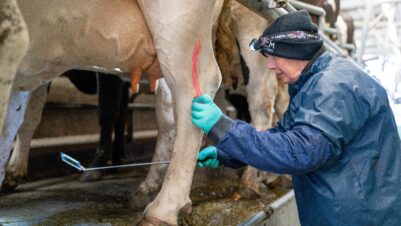
Q fever is an infectious disease caused by an intracellular bacterium, Coxiella burnetii, which can infect a wide range of hosts and survive for long periods in the environment (Welsh et al., 1959; Hawker et al., 1998; McCaul and Williams, 1981). Q fever is also a zoonotic disease and can be a health risk for vets, farmers and other workers in the livestock industry (Figure 1) (Maurin and Raoult, 1999).
Although C. burnetii can infect different hosts, ruminants play a significant role as the main reservoir of human infection. Among ruminants, goats show the most acute clinical signs. However, many studies have highlighted how Q fever is widely present in dairy farms and can affect the health and productivity of cattle (Barberio et al., 2017).

Prevalence of Q fever
According to recent studies examining the presence of the bacterium in bulk-tank milk (BTM) across Europe, around half of dairy herds are exposed to Q fever (Barberio et al., 2017). The exposure to Q fever is also high for goats, with one out of three herds considered at risk (Guatteo et al., 2011).
A study […] has highlighted that out of 155 herds tested with a PCR test, 118 tested positive for C. burnetii, demonstrating active Q fever on-farm
In Great Britain, the seroprevalence of C. burnetii in cattle has been established to be as high as 79 percent, according to the most recent study on endemic diseases (Velasova et al., 2017). Further, a study in dairy herds in the southwest of England has highlighted that out of 155 herds tested with a PCR test, 118 tested positive for C. burnetii, demonstrating active Q fever on-farm (Valergakis et al., 2012).
Transmission
C. burnetii is highly resistant in the environment and can be transported by the wind for up to 11 miles (Hawker et al., 1998). Thus, inhalation of aerosols containing bacteria represents the main route of infection in cattle (Welsh, 1957). Historically, Q fever had been considered a tick-borne disease, and although ticks are not the main transmission route, C. burnetti can live in ticks for years with scope for transdermal infection (Heinzen et al., 1999; To et al., 1995).

One of the biggest challenges with Q fever transmission is environmental contamination (Welsh et al., 1959). C. burnetii is shed mainly via birth fluids and the placenta in the periparturient period. However, in cattle, shedding may also occur through vaginal mucus, milk, faeces and urine from cows at any stage of lactation (Figure 2) (Guatteo et al., 2006, 2007). For this reason, although calving and lambing present a higher risk, shedding from animals in the lactating stage may still be possible, increasing the risk of environmental contamination and making biocontainment very challenging.
Although calving and lambing present a higher risk, shedding from animals in the lactating stage may still be possible, increasing the risk of environmental contamination and making biocontainment very challenging
Q fever in humans
Q fever is a zoonosis that typically manifests with acute flu-like symptoms in humans. Signs usually regress spontaneously within a few days. However, in 4 percent of cases complications including pneumonia, hepatitis encephalitis and meningitis may develop, requiring hospitalisation (Dragan and Voth, 2020). The disease can also develop chronically in 2 percent of cases, leading to cardiac complications or chronic fatigue (Dragan and Voth, 2020). In pregnant women, it can lead to abortion, premature birth or foetal death (Arricau-Bouvery and Rodolakis, 2005).

Clinical presentations
In experimental infections in heifers, two clinical phases have been identified: acute and chronic. In the acute phase, severe hyperthermia and rapid pneumonia were observed, with spontaneous clinical recovery in the following seven days. In the chronic phase, the main clinical signs were abortions and infertility (Plommet et al., 1973).
Infected dairy cattle generally do not display obvious clinical signs and the clinical presentations are related to the chronic nature of the disease. Nevertheless, there is evidence that Q fever is implicated in reproductive disorders, such as abortions. In one study, C. burnetii was identified directly in the macrophages of the endometrium of cows with poor fertility (repeat breeding), establishing a direct link between poor fertility and endometrial lesions due to Q fever (Figure 3) (De Biase et al., 2018).
Furthermore, there is evidence of Q fever being linked to poor conception at first service, embryonic loss and irregular return to heat (Dobos et al., 2020). Other studies have found a correlation between Q fever and a higher risk of retaining the placenta (Ordronneau, 2012). Lastly, in an Italian study, positive Q fever PCR tests on BTM correlated with a higher incidence of metritis and clinical endometritis (Valla et al., 2014).
In goats, the clinical picture is different. Q fever is one of the main abortive infectious agents responsible for abortion storms that can affect up to 90 percent of pregnant animals (Agerholm, 2013). Milk production of the small ruminant dairy breed may also be affected by the presence of C. burnetii. In Australia, scientists have found that infected goats produced 20 percent less milk than goats free from the disease (Canevari et al., 2018).

When to suspect Q fever
Due to the nature of Q fever and the fact it is similar to many other diseases in dairy herds, diagnosing Q fever can be challenging. Unexplained abortions, pregnancy loss at any stage or stillbirth, high levels of metritis and endometritis, or unexplained poor fertility performance (eg repeat breeding, higher calving to conception rate, embryo loss) must trigger a Q fever investigation (Figure 4).
Unexplained abortions, pregnancy loss at any stage or stillbirth, high levels of metritis and endometritis, or unexplained poor fertility performance must trigger a Q fever investigation
Diagnostic tools
The diagnostics should combine direct methods of identification of the bacterium, such as PCR in the BTM, and a serological approach, such as an ELISA from the blood serum of cows with reproductive disorders. If the BTM is positive for C. burnetii or more than half of the problematic cows sampled are seropositive, then the bacterium is circulating in the herd and is likely to play a role in the problems observed.
Chronic abortions in cattle
In cases where there has been a series of abortions, the correct approach is to rely on direct identification via PCR on vaginal samples on at least two aborted dams. It is essential that the sampling is done within seven days following the abortion and the sample is sent to a veterinary diagnostic centre immediately. The placenta and stomach content of aborted calves can also be sent as sample materials for investigation.
In cases where only one PCR is positive, it is beneficial to collect blood from cows that were recently aborted or with reproductive disorders to assess seroprevalence. If two PCR tests come back positive, or more than half of the cows are seropositive, the chances that Q fever is responsible for the abortions are very high.
For small ruminant abortion, it may be economically viable to run a PCR analysis on a pool of vaginal samples collected from three freshly aborted ewes or does. If the pool is positive, Q fever is likely to be the cause of the recent abortions.
How to control the disease
The Q fever control strategy involves combining biosecurity measures and vaccination.
Biosecurity
Biosecurity measures can help reduce or prevent exposure to contaminated aerosols and minimise environmental contamination. Such measures may include strict hygiene during calving or lambing, and avoiding spreading manure, especially in certain weather conditions (eg strong winds) (Tissot-Dupont et al., 2004). However, the most critical biocontainment measures for Q fever are systematically removing and destroying the placenta and aborted foetus, alongside cleaning the calving area.
The most critical biocontainment measures for Q fever are systematically removing and destroying the placenta and aborted foetus, alongside cleaning the calving area
Vaccination
The cornerstone of Q fever control is vaccination. A phase I vaccine is available for cattle and goats, the main advantages of which are a significant decrease in the excretion of C. burnetii in a farm environment (Pinero et al., 2014; Guatteo et al., 2008), an improvement in fertility and a decrease in the abortion rates in dairy cattle (Lopez Helguera, 2014). The vaccine can also be safely used in pregnant animals and can help to minimise shedding from infected cows, protecting naive animals.
What about antibiotics?
In human medicine, Q fever is treated with the use of antibiotics. However, antibiotics (such as oxytetracyclines) cannot prevent the disease or decrease bacterium excretion from infected animals. Thus, treatment with antibiotics has little effect on disease control in cattle (Taurel et al., 2012).
Costs associated with active Q fever
Quantifying the costs associated with Q fever in dairy farms is challenging, and they must be considered on a case-by-case basis. However, they can be summarised in:
- Direct and indirect costs of retained placenta/metritis/endometritis
- Fertility key performance indicators, including:
- Calving to conception interval (Figure 5)
- Embryo loss
- Culling due to fertility issues
- Abortion rate

Summary
Despite historical underestimations, C. burnetii is highly prevalent in UK dairy herds and can survive for a long time in the environment, presenting Q fever as a significant problem to dairy herds and as a threat to human health. The real impact of the disease must be investigated further, especially considering the studies linking C. burnetti to fertility issues and other reproductive disorders that can impact dairy cows’ health and performance. Understanding clinical presentations, diagnosis and the true impact of the disease on farm performance is the cornerstone for practitioners to help farmers develop a control strategy and improve productivity.


