
Since the Pet Travel Scheme (PETS) was relaxed in 2012, pet travel has increased year on year at a time of increased human migration, pet movement and climate change, providing favourable conditions for the rapid spread of parasites and their vectors. This has increased the risk of pets and their owners encountering exotic parasites while abroad, and their introduction to the UK through travelled dogs and cats.
Legal and illegal importation of dogs from foreign countries are also increasing the risk of foreign parasite and vector introduction,with Defra reporting 30,000 dogs being imported into the UK in 2016 alone. As a result, a variety of exotic parasites are being seen in UK practices on a regular basis.
As well as microscopic vector-borne pathogens such as Ehrlichia and Leishmania, exotic worms are also being seen with increasing regularity. Veterinary professionals need to be aware of these foreign travellers to maintain animal and human health as well as national biosecurity. There are three exotic parasitic worms (and one worm-like parasite) that are likely to be coming to your practice in the near future.
Thelazia callipaeda
Thelazia callipaeda, also known as the “oriental eye worm” due to its high incidence in Asia, is a zoonotic vector-borne nematode which resides in the conjunctival sac of definitive hosts such as carnivores, rabbits and humans. The worm has been spreading through Europe in recent years, following its fruit fly vector, Phortica variagata. The fly transmits the parasite when feeding on lacrimal secretions. Although often non-pathogenic,infection can commonly cause conjunctivitis, keratitis, ephiphora, eyelid oedema, corneal ulceration and, in serious cases, blindness.
Since T. callipaeda was first reported in Italy in 2007, autochthonous cases have spread east and northwards, being reported in France, Germany, Switzerland, Spain and Portugal, and more recently into eastern Europe (Mihalca et al., 2015).The first confirmed cases in the UK were recently recorded in dogs imported from Romania, Italy and France. P. variegate, the primary fruit fly vector of T. callipaeda, has been recorded in the UK with conditions favourable for spread (Graham-Brown et al., 2017). It is vital therefore that infections are detected rapidly in travelled dogs and treated.
Diagnosis can be achieved by visualisation of the worms on the conjunctiva of the eye. Sedation maybe required to fully visualise worms residing under the nictitating membrane, where many or all the worms will be residing. This should be carried out on any dog with travel history abroad with conjunctivitis or other eye surface pathology of unknown cause. Elimination of the worms can be achieved by flushing of the eye in conjunction with a systemic macrocyclic lactone. An Imidacloprid/moxidectin spot on treatment or two milbemycin oxime oral treatments given two weeks apart are both licensed for treatment.
.png)
Heartworm (Dirofilarial immitis)
Dirofilaria immitisis a filarial heartworm primarily of dogs, but also can infect ferrets and felines. It is a significant cause of heart disease and also of bronchitis in cats. Transmission occurs through feeding by infected mosquitoes. Infections may remain subclinicalor potentially severe signs can develop. Acute clinical signs include:sudden death, anorexia, weakness, dyspnoea, ascites, anaemia, haemoglobinuria, vomiting and, rarely, pleural effusion. Chronic signs are due to inflammatory responses and aberrant migration. These tend to be respiratory in nature and include:coughing, dyspnoea, anorexia and vomiting.
It is present throughout Southern Europe but the mosquitoes capable of transmitting the parasite can be found throughout the whole of Europe, including the UK. The climate in northern and central Europe has been too cold to allow D. immitis to complete its life cycle in the mosquito,but climate change in combination with increased movement of pets and people throughout Europe is allowing the parasite to spread. The distribution of D. immitis is illustrated in Figure 1, and it is now endemic in eastern European countries including Serbia, Romania and Bulgaria.
Rapid diagnosis in imported pets is important to manage infection and preventative treatment for pets travelling to endemic countries vital to keep pets safe. A number of useful diagnostic tests are available in suspected cases.
Examination of blood for microfilaria
Concentration techniques, such as Knott’s test (a form of sedimentation test) and buffy coat examination,are more sensitive than direct whole blood examination in canine patients, but still extremely insensitive in the cat due to microfilariae being absent or present in low numbers.
Radiology
Thoracic radiographic changes are not pathognomonic but diffuse interstitial patterns, right sided heart enlargement and enlargement of the pulmonary arteries, particularly the caudal lobar arteries may all be present.
Ultrasound examination
The cuticles of adult heart worms are highly echogenic and so in experienced hands echocardiography is very sensitive and specific.
Antigen serology
This test is considered the gold standard in the living canine patient. It is highly specific and in canine patients, also highly sensitive. Although specificity still approaches 100 percent in cats, it is much less sensitive. Sensitivity increases as adult female worm burden increases with the test detecting antigens in uterine secretions.
Antibody serology
This is the most sensitive blood test available for heartworm diagnosis in cats (Atkins, 1999). A positive result may indicate past or present exposure to infection and should be interpreted in relation to clinical signs and other diagnostic tests.
.jpeg)
Treatment of infections with adult worms requires surgical removal or treatment with an adulticide. Intravascular snares and forceps are often used in endemic countries to remove large numbers of worms in the least invasive manner, and this is essential in caval syndrome, where death can occur within two days of onset of acute signs.
Echocardiographic visualisation of large numbers of worms in the pulmonary artery allows the use of flexible alligator forceps under fluoroscopic guidance to remove the worms (Figure 2). These techniques require experience and specialised equipment however, and as a result, most UK vets will require a medical approach to treatment of adult worms.
The vital components of any treatment protocol are pre-treatment with doxycycline and a macrocyclic lactone prior to three adulticide treatments. Variation in protocols tends to occur around the length of these pretreatment periods, but all agree that three adulticide treatments are essential to maximise elimination of adult worms. These are given by deep intramuscular injection, two being given 24 hours apart, 30 days after administration of the first injection. An example of a typical adulticide treatment programme is:
- Day 1: Doxycycline 10mg/kg sid orally for 30 days
- Heartworm preventative (macrocyclic lactone on day 0 and 15)
- Day 30: Melarsomine dihydrochloride 2.5mg/kg IM
- Day 60 and 61: Melarsomine dihydrochloride 2.5mg/kg IM
The patient should then be tested for microfilariae 30 days post-treatment and antigen serology tested six months post-treatment. Prednisone has some benefit in reducing the risk of thromboembolic complications if given alongside adulticide treatment where worm burdens are high. If high burdens are suspected, then oral prednisolone can be used from the initiation of adulticide treatment at 0.5mg/kg twice a day for one week and 0.5mg/kg once daily for the second week, followed by 0.5mg/kg every other day for two weeks. These doses are also useful in managing bronchitic signs.
The three most significant factors involved in post-adulticide treatment complications are the severity of existing pulmonary vascular disease, the number of worms present and level of exercise. Of these three, over-exercise is thought to be the most significant. Exercise should therefore be restricted during treatment, starting from day 0 to at least one month after the last adulticide injection. The highest risk period of complications from pulmonary thromboembolism is 7 to 10 days after adulticide treatment, but can occur up to four weeks after adulticide treatment is completed.
An alternative treatment which avoids the use of the adulticide melarsomine is the “slow kill” regime. This should not be used as a first-choice treatment as it has been linked to the development of resistance (Bowman et al., 2012) and carries some risk of anaphylaxis due to the killing of microfilariae from active infection over a long period of time. This is particularly true when large numbers of micro-filariae are present in the circulation. It should therefore only be used if surgery is not indicated or practical and melarsomine is not available. The protocol is:
- Doxycycline 10 to 20mg/kg sid or bid for 30 days
- Ivermectin at minimum dosage of 6 to 12mcg/kg bimonthly or topical moxidectin (2.5mg/kg) bimonthly until two consecutive negative antigen test results after 12 months
Exercise restriction is also recommended with this protocol from the start of treatment until infection is eliminated.
.JPG)
Dirofilaria repens
Dirofilaria repens is closely related to D. immitis but is less pathogenic to cats and dogs with adult worms living in the skin. It has the potential to cause creeping eruptions and conjunctivitis in people. It can complete its life cycle at lower temperatures than D. immitis (Morgan, 2016) and so has a far wider distribution across Europe (Figure 1).
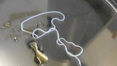
The first cases confirmed in the UK were from dogs imported from Corfu and Romania (Agapito et al., 2017; Wright, 2017); if infected dogs continue to enter the UK and are not treated quickly, UK mosquito populations could be exposed to the parasite and the disease could become endemic. Imported dogs should be checked for ocular and skin lesions and diagnosis confirmed by Knott’s test for microfilaria, biopsy or identification of aberrant adult worms (Figure 3). These are long and thin without any grossly obvious identifying features. Males are about 5 to 7cm and females 10 to 17cm in length. Under magnification, a series of small papillae arranged in a “v” shape may be seen in male worms. They have two unequal spicules at their caudal end.
Discrete subcutaneous nodules containing adult worms can be removed surgically with an excellent prognosis. Moxidectin/imidacloprid spot on preparations are licensed for medical treatment.
.JPG)
Linguatula serrata
This parasite is known as a “tongue worm”, but is actually a pentastomid, more closely related to arthropods than true worms. The adult parasite is an elongated tongue-shape (Figure 4) and is found in the nasal cavities or sinuses of dogs and foxes. Infection occurs through the ingestion of nymphs in raw offal of infected intermediate hosts, such as ruminants, rabbits and horses. Eggs from the adult parasite are passed in the faeces or nasal secretions of infected dogs and are immediately infective. Adult parasites are large, with females typically 30 to 130mm in length.
Although the parasite has been reported in UK foxes, it is thought to be rare. There has, however, been a sharp in-crease in clinical cases reported in UK dogs imported from eastern Europe and the Middle East where raw meat is routinely fed. This is a concern due to the zoonotic potential of the parasite. Although in endemic countries zoonotic infection occurs primarily through the ingestion of raw or undercooked viscera, it can also occur through ingestion of eggs in the environment or in mucoid discharge from infected dogs’ noses. This can lead to a variety of clinical presentations, including nasopharyngitis, blocked nasal passages, visceral pain and aberrant larval migration to the anterior chamber of the eye.
Imported dogs expulsing adult parasites or presenting with rhinitis, gagging or chronic upper airway signs should be examined by endoscopy to check for infection. Alternatively, nasal discharge can be tested by flotation methods for eggs. This can also be performed on faeces but with a much lower sensitivity. There are reports of infections being successfully treated with oral milbemycin or moxidectin/imidacloprid spot on solution. Parasites may also be phys-ically removed by endoscopy. Infections should be treated promptly and good hand hygiene maintained by owners.
Exotic roundworm prevention
Pets travelling to D. immitis endemic counties should have monthly treatment with a licensed macrocylic lactone for heartworm prevention. Moxidectin/imidacloprid spot on solutions are also licensed for D. repens prevention. While there is no specific preventative treatment for T. callipaeda infection, routine macrocyclic lactone treatment may be of some benefit. A licensed pyrethroid fly repellent will also have some effect in the reduction of vector-borne disease.
ESCCAP UK and Ireland recommends four key steps (the “four pillars”) when dealing with all imported or travelled pets arriving in the UK to manage exotic parasite risk:
- Check for ticks and identify any found.
- Treat dogs again with praziquantel within 30 days of return to the UK and treat for ticks if treatment is not already in place.
- Recognise clinical signs relevant to diseases in the countries visited or country of origin.
- Screen for Leishmania spp., heartworm and exotic tick-borne diseases in imported dogs.
Following the “four pillars” allows veterinary professionals to prepare owners if parasites are present, improve prognosis of clinical cases, minimise the risk of spread of any dis-ease and carry out effective disease/parasite surveillance.
Conclusion
Increased movement of pets and fluid vector and parasite distributions in Europe make protecting UK pets and owners from exotic disease increasingly challenging. Exotic par-asitic worms and worm-like organisms must be considered as possible infections in imported and inadequately protected travelled pets. Vets and nurses are in the front line of UK biosecurity and must be prepared to give accurate travel advice to clients and remain up to date with the latest parasite distributions in Europe and across the globe.