Veterinarians have long been aware that common companion animals carry zoonotic diseases, and exotic pets are no exception.
Zoonotic diseases are diseases that can pass from animals to people, resulting in symptomatic or asymptomatic infections. They are not uncommon, and it has been postulated that up to 75 percent of emerging human diseases are of zoonotic origin (Taylor et al., 2001). The list of zoonotic diseases carried by exotic pets is extensive, and veterinarians should ensure that they are aware of species-specific zoonoses when treating exotic patients.
This article discusses some of the most common zoonoses a clinician may encounter in practice but it is by no means exhaustive.
Psittacosis
Psittacosis is a human disease syndrome resulting from an infection with the bacteria Chlamydia psittaci. The disease can also be known as ornithosis, parrot fever or chlamydiosis (Balsamo et al., 2017), but is commonly known as psittacosis due to how well associated it is with psittacine birds (Crosta et al., 2016).
The causative agent is an obligate intracellular bacterium usually transmitted following inhalation of infectious organisms from respiratory secretions, feather dander or dried faeces, or direct beak-to-mouth contact between a bird and human (Zwart, 2016). This is particularly important for owners who engage in over-amorous behaviour with their pet parrots, including mouth-to-beak contact when kissing their birds on the head, beak or over the nares.
Common carriers of C. psittaci include psittacines (parrots), poultry, doves, pigeons and birds of prey (Balsamo et al., 2017). Transmission of C. psittaci between humans is rare but has been reported (Hughes et al., 1997; McGuigan et al., 2012).
Clinical signs

Clinical signs in humans can be absent, but usually, there is an incubation period of 5 to 14 days following exposure, although incubation times as long as one month have been observed (Crosta et al., 2016).
Symptoms are individual but range from a mild, non-specific malaise to pneumonia and systemic disease, with mortalities being reported (Balsamo et al., 2017). Commonly reported symptoms include fever, chills, muscle aches, dry cough, headache and upper respiratory tract signs (Crosta et al., 2016).
Psittacosis should be strongly suspected in people who have contact with birds and are showing clinical signs, regardless of whether the bird or birds are showing clinical signs. Clinical signs in birds can be non-specific, such as lethargy, hyporexia or anorexia, regurgitation, green faeces or conjunctivitis, or they can manifest as respiratory disease with obvious dyspnoea and oculonasal discharge (Figure 1) (Zwart, 2016).
Prevention and treatment
There are set criteria that define suspected cases, probable cases and confirmed cases in avian patients, and probable or confirmed cases in humans (Table 1).
In humans, the treatment of choice is tetracyclines, with most clinical cases treated with oral doxycycline (Balsamo et al., 2017). This is similar to treatment of avian patients, where oral tetracyclines are also recommended. It is essential that the treating clinician discusses the potential for zoonoses with the owner if psittacosis is suspected or diagnosed in any avian patient. This is especially crucial if immunosuppressed people are living in the home with the bird or have come in contact with the bird.
C. psittaci is very treatable in avian and human patients, and reinfection can be avoided by following quarantine protocols after treatment. Confusion between psittacosis and other diseases may cause human medical professionals to recommend removing or rehoming the bird, but this is rarely, if ever, necessary.
| Avian patients | Human patients | ||
|---|---|---|---|
| Suspected cases: one of these criteria | Compatible illness not confirmed by a laboratory but is epidemiologically linked to a confirmed case in an avian or human patient A patient with no clinical signs but a high serological titre or detection of chlamydial antigen Compatible clinical signs with a positive result from a non-standardised test Compatible clinical signs that respond to therapy | n/a | |
| Probable cases: compatible clinical signs and one of these criteria | A single high serological titre following onset of clinical signs Chlamydial antigen detected in respiratory secretions, faeces or cloacal swab | Probable case: compatible clinical signs and one of these two laboratory results | An IgM titre below 32 in at least one serum sample obtained after onset of clinical signs Detection of C. psittaci in a respiratory specimen (sputum, pleural fluid or tissue) via PCR |
| Confirmed cases: one of these criteria | Isolation of C. psittaci from a clinical specimen Identification of antigens in the patient’s tissues with immunofluorescence A fourfold or greater increase in serological titres taken a minimum of two weeks apart and assayed simultaneously at the same laboratory Identification of C. psittaci within macrophages in smears or tissue using Macchiavello or Gimenez stain | Confirmed cases: compatible clinical signs and one of the following laboratory results | Isolation of C. psittaci from respiratory secretions or blood Fourfold or greater increase in serum IgG by use of complement fixation or microimmunofluoresence between samples obtained two to four weeks apart |
Salmonellosis
Historically, salmonellosis has been associated with reptiles and reptile ownership, but this association has also evolved to include amphibians (Souza, 2011). While reptiles are known to carry Salmonella, with up to 93.7 percent of reptiles harbouring Salmonella spp. as part of their normal gastrointestinal flora (Sodagari et al., 2020), the majority of reptiles are asymptomatic carriers. In fact, Salmonella infection from reptiles is rare. A 2004 study in the United States attributed only 6 percent of salmonellosis cases to contact with reptiles or amphibians (Mermin et al., 2004). Most salmonellosis cases are attributed to contaminated food, most commonly of poultry origin such as eggs and meat (Marin et al., 2021).
The biggest concern for infection with Salmonella spp. is the increasing number of antibiotic-resistant strains, which is a public health concern. Multidrug-resistant Salmonella strains have been identified in reptiles (Wei et al., 2019) and there is concern that, as Europe is a large-scale exporter of many reptile species, the use of antibiotics by commercial breeders and exporters is promoting strains of antimicrobial-resistant Salmonella (Marin et al., 2021).
Reptile-to-human transmission of Salmonella spp. is via indirect or direct contact with the patient or their faeces (Sodagari et al., 2020). Those most at risk include children, whose immune systems are still developing and potentially have poorer hygiene than adults, and immunosuppressed people, such as the elderly.
Clinical signs
Clinical signs in humans are often only self-limiting gastroenteritis with fever, diarrhoea, nausea, vomiting and abdominal pain, but death can occur with severe illness in immunocompromised individuals (Marin et al., 2021).
Prevention and treatment

The best way to prevent Salmonella infection is good hygiene. Always wash hands before and after handling any reptile patients and their faeces. Clinicians and veterinary staff should always wear gloves when handling faeces from any species but should also consider wearing gloves when handling reptiles in any context (Figure 2). Clients should be discouraged from kissing reptiles or having them close to their face. Reptiles should not be allowed in areas where their owners are eating or preparing food. In addition, a separate sink, such as a utility sink, is recommended to wash reptile bowls and enclosure furniture (Sodagari et al., 2020).
The use of antibiotics to treat reptile patients whose faeces has positively cultured Salmonella spp. is controversial. Treatment of healthy reptiles carrying Salmonella spp. is unlikely to clear the bacteria and can result in the development of antibiotic-resistant strains (Marin et al., 2021). In reptile cases with gastrointestinal signs, assessment of any other possible underlying causes, such as endoparasites, should be investigated, and supportive care should be considered prior to antibiotics.
Dermatophytosis

Dermatophytosis, or ringworm, is a common presentation in small mammal species, whether exotic pets or British wildlife. The most commonly affected species is guinea pigs, with infection often occurring in younger animals with less competent immune systems (Longley, 2009). Dermatophytosis has also been reported in rats, mice, hamsters, hedgehogs (Figure 3), chinchillas and ferrets (Pollock, 2011).
Dermatophytosis in small mammals is commonly caused by Trichophyton mentagrophytes, although other Trichophyton species have been implicated and saprophytic species (such as Microsporum spp.) have also been reported (Pollock, 2011). Infection is usually caused by immunosuppression, with several factors, including overcrowding, dietary deficiencies, stress, ectoparasites and pregnancy, postulated to increase the risks of infection (Kraemer et al., 2012).
Clinical signs
Clinical signs in guinea pigs and other small mammals include non-pruritic crusting or scaling around the nose, eyes and ears, although immunocompetent animals may be asymptomatic carriers (Longley, 2009).
Infection in humans is caused by direct contact, with one study finding 25 percent of dermatophytosis cases in guinea pigs had zoonotic spread to humans and that children were at a higher risk for contracting dermatophytosis than adults (Kraemer et al., 2012). In humans, the face, neck and arms are the most common sites for lesions which develop as raised, red, pruritic lesions (Kraemer et al., 2012).
Diagnosis can be made by the presence of characteristic clinical signs or fungal culture (Longley, 2009).
Prevention and treatment
Treatment for dermatophytosis involves systemic or topical anti-fungal medication. Consideration should be given that most mammals are fastidious groomers, thus topical preparations may be ingested during routine grooming.
The environment should also be disinfected to prevent reinfection of the skin, and any underlying predisposing factors should be corrected. Environmental decontamination with a 1:10 bleach solution should be sufficient (Longley, 2009).
Keeping veterinary professionals safe

It is important that veterinary staff working with exotic pets are aware of the potential zoonotic diseases and take precautions to avoid transmission.
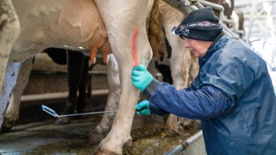
As with all diseases, good hygiene is the mainstay of preventing the spread of infection. The use of gloves and other personal protective equipment (PPE) when handling exotic pets is recommended, with consideration for the level of PPE required assessed on a case-by-case basis (Figure 4). Hand washing and sanitising surfaces such as examination tables and cages between patients is essential in minimising the spread of infectious diseases to other patients and to staff working within the veterinary clinic.
Education of owners is also paramount, particularly with owners who express over-amorous behaviour towards pets with potential zoonotic disease. Kissing or face contact with any exotic pet is never recommended and should, ideally, be completely avoided.
Conclusion
Clinical staff should be educated and aware of potential zoonotic risks in all patients presenting to their clinic. It is rare that zoonotic disease is contracted by veterinary staff or owners, but care must be taken with immunocompromised people or patients. A thorough understanding of zoonotic diseases is important to educate clients so that they are aware of the risks for themselves and family members, should a zoonotic disease be diagnosed in their pet. Good hygiene is the mainstay of preventing spread of zoonotic disease.