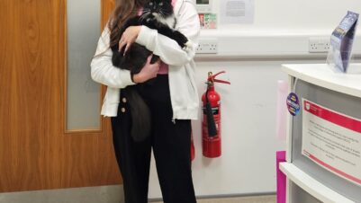
New research, led by orthopaedic Specialist Steve Bright at Manchester Veterinary Specialists, has evaluated the use of a 3D-printed patient-specific guide for the treatment of distal tibial varus deformity in Dachshunds.
The study found 3D-printed patient-specific osteotomy and reduction guide systems facilitated the accurate correction of tibial varus deformity with very good clinical outcomes. It also found an opening osteotomy, stabilised with orthogonal locking plates and without the application of bone graft, led to satisfactory bone healing in all cases.
Pes varus is a bone deformity where the distal tibia is turned inward toward the body. Most affected Dachshunds are juveniles with no history of trauma, suggesting a genetic predisposition. Typically, affected dogs develop gait abnormality, with an abnormal posture and a “bow-legged” appearance.
Surgical options to correct tibial malalignment have traditionally relied on 2D radiographic planning – to identify and quantify the correction needed and to plan the position and orientation of the osteotomy. Whilst excellent outcomes can have been achieved this approach has potential drawbacks. Accurate quantification of tibial torsional can be challenging from 2D radiographs alone, which is where 3D assessment via CT becomes invaluable.
The Surgical correction of pes varus deformity in dachshunds using three dimensional-printed patient-specific guide system: nine tibiae in seven cases study evaluated the accuracy of a pes varus deformity correction in Dachshunds using CAD-based planning, 3D-PSGSs and orthogonal locking plate stabilisation.
Data from computed tomography were processed to obtain virtual 3D-models of the tibias, which were used for; a 3D-printed patient-specific guide system design; computer aided design-based surgical planning; and, evaluation of planned versus achieved tibial correction. Surgery, comprising an opening osteotomy, stabilised with orthogonal locking plates and without the application of bone graft, was then conducted. Radiographic and CT examination of the pelvic limb was performed immediately post-operatively, and radiographs and CT were obtained at follow-up examinations. Clinical outcomes were evaluated by lameness assessment and post-operative owner-reported questionnaire at a minimum of 15 months.
All dogs went on to achieve a very satisfactory clinical outcome. A range of motion was considered within normal limits in all tarsi, none of which appeared uncomfortable during manipulation. There was excellent correlation between planned and achieved deformity correction, with mean translational error <1mm in all planes, and mean angulation correction error <2mm in all planes. In follow-up radiography in 75 percent of the tibiae in the study, 75 percent cortical bone healing was noted by 6 to 8 weeks post-operatively and marked osseous infilling was present (note, one owner declined). In four cases imaged over 10 weeks post-operatively, osseous infilling was complete. All owners also reported a very satisfactory post-operative outcome.
Steve Bright, clinical director and specialist in small animal orthopaedics at Manchester Veterinary Specialists, said: “Two-dimensional derived reference values are intrinsically inaccurate when applied to a complex 3D object such as a multiplanar bone deformity.
“Our collaborative study, in conjunction with our colleagues at Vet3D, demonstrated that CAD-based surgical planning and the use of 3D-printed patient-specific osteotomy and reduction guides resulted in accurate tibial correction and good post-operative tibial conformation and limb function. Orthogonal, double plating with locking plates surgery was associated with rapid bone healing within the osteotomy gap, which did not require any form of bone graft, and was associated with excellent clinical outcomes.
“We feel this this type of surgical planning and technique has the potential to significantly improve our accuracy in the management of this condition.”