Human intervention is often unnecessary following the birth of a healthy puppy or kitten to an unafflicted dam. Maternal instincts are sufficient to revive a vigorous newborn, which this article will define as a neonate in the first hours of life. Assistance and resuscitation are often required following dystocia and always required following caesarean section. Such resuscitation is regularly performed by nursing and lay staff with minimal formal training in how to optimise outcome, particularly for those newborns in extremis.
Optimising newborn survival
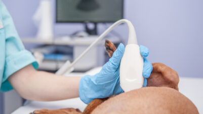
Newborn survival is maximised by ensuring dystocia is promptly recognised and treated. We can achieve this by fine-tuning the continuum of clinical decisions from telephone triage to the clinic and operating theatre if necessary. Delays or poor decision making may result in non-vigorous newborns or the loss of an entire litter. Table 1 lists the tele-triage situations where veterinary assessment is indicated alongside the indications for caesarean section during assessment.
| Indications for triage | Indications for caesarean section |
|---|---|
| Dam over term: over 66 days from luteinising hormone surge (if the date is known) or over 72 days since the last mating Failing to progress into labour 24 hours after a documented temperature drop If parturition lasts longer than 24 hours in a dog or 36 hours in a cat If the neonate is not born within four hours after “first waters broke” A period longer than four hours between neonates (except cats) Yelping or biting flank, hindquarters or vulva while straining More than 30 minutes of intense unproductive straining or strong abdominal contractions Increasing volumes of uteroverdin discharge prior to the neonate being born. (This discharge is normal in the 30 minutes before delivery but if volumes are noticed without successful delivery within 30 to 60 minutes, this is an emergency as excessive/increasing amounts suggest placental separation and, therefore, loss of foetal blood supply) If part of the foetus or membranes are visible and not being expelled Excessive haemorrhage Collapsed or exhausted dam Stillbirth – suggests dystocia and problems with litter Foul-smelling discharge | Persistent foetal distress diagnosed via ultrasound Foetus stuck in the birth canal with manual manipulation failing Uterine inertia unresponsive to medical management Excessive uteroverdin passed without neonate born Excessive blood loss Known foetal oversize Large litter precluding medical management Foetal death |
Effective resuscitation requires adequate equipment (Box 1) and staff numbers, the latter of which can be challenging in some clinical environments. Preparation of staff and equipment prior to intervention can both be lifesaving and reduce stress for all involved.
The major influences on neonatal vigour following caesarean section are hypoxia and the effect of depressant drugs, such as opioids, used during surgery. Specific drug protocols and their effect on dam or litter is beyond the scope of this article.
| Oxygen source(s) Numerous warm dry towels, air heaters and/or “hot hands” Small masks with a diaphragm Laryngoscope with a small blade Gauze swaps Suction bulbs and cotton buds Doppler Small endotracheal (ET) tubes and extra 3F ET adapters All sizes of IV catheter – 14 to 16G IV catheters for intubations, 22 to 26G IV catheters for jugular access and umbilical vein catherterisation Needles – 21 to 23G for intraosseous access. Spinal needles are preferable Syringes Glucose Warmed crystalloid fluids |
The tenets of neonatal resuscitation
As in other areas of CPR, the tenets of neonatal resuscitation follow the mnemonic “ABC”: airway, breathing and circulation.
Airway

During caesarean section, the surgeon should avoid the removal of the foetoplacental unit due to the risk of foetal anoxia (Onclin and Verstegen, 2014). The amniotic sac should be opened, and the umbilical cord clamped and dissected. The newborn should then be aseptically handed to a colleague. The author recommends leaving an umbilical cord long enough to allow umbilical catheterisation if necessary. Foetal membranes and fluid should then be cleared from the nares and mouth using a bulb syringe (Figure 1) or DeLee suction catheter. Mechanical suction should be avoided due to the potential trauma of delicate neonatal tissue and the risk of vagal stimulation and laryngeal spasm (McMichael and Dhupa, 2000).
Tilting the neonates so the hindquarters are slightly raised and vigorously rubbing them with a warm dry towel provides tactile stimulation to encourage spontaneous breathing while warming, drying and aiding airway clearance. Rubbing the fur “against the grain” in the lumbar area stimulates crying and helps airway clearance. Swinging is contraindicated due to the risk of intracranial haemorrhage, traumatic brain injury and gastric fluid aspiration (Traas, 2008).
Breathing
Hypoxia
Hypoxia of a foetus or newborn may be common in dystocia cases due to:
- Relative hypoxia of a dam
- Poor placental blood flow – this can be caused by inappropriate oxytocin doses, which cause uterine tetany (Davidson, 2001), and hypothermia during anaesthesia. Furthermore, states of pain, stress, hyperventilation and hypotension in the dam all reduce uterine and placental blood flow
- Traction on the umbilical cord during delivery (Münnich and Küchenmeister, 2014)
- Occlusion of the umbilical cord during delivery (Kuttan, 2018)
- Placental separation without the subsequent delivery of neonate
- Foetal/neonatal hypothermia
- Asphyxiation on foetal fluids (more common in “breech” presentation) or meconium
Unique to neonates, systemic hypoxia manifests as bradycardia, which is thought to be caused by myocardial hypoxia and may act as a protective mechanism which results in longer survival times in low-oxygen states. Neonates spontaneously breathing with a heart rate of over 120 to 150bpm immediately following delivery are likely to be stable and require minimal assistance following initial airway clearance. The heart rate will likely rise to a normal neonatal rate of around 200bpm (Moon et al., 2001).
While routine oxygen administration to vigorous newborns may be deleterious, those that are cyanotic or bradycardic should receive flow-by oxygen while towel drying (Kattwinkel et al., 2010). Multiple oxygen sources may be required to resuscitate multiple struggling newborns.
Gasping and apnoea
Gasping or apnoea is common in a non-vigorous newborn. Aiding ventilation and reversing apnoea are the primary focus of resuscitation in these patients. Breathing can be encouraged with tactile stimulation of the genital and umbilical areas (Fox, 1964). Acupuncture of the Jen Chung (GV 26) point in the nasal philtrum has also been described as a respiratory stimulant. This is performed by piercing the philtrum with a 25G hypodermic needle and rotating it once bone or cartilage is contacted (Skarda, 1999).
Medication
Any opioids administered to the dam prior to delivery will flow through the placenta and depress the foetus. If necessary, reversal of opioids with naloxone can encourage breathing and vigour during resuscitation. The author would not advise using opioids in the pre-delivery anaesthetic plan unless naloxone is stocked and available for subsequent newborn resuscitation.
The appropriate use of the central respiratory stimulant doxapram (Dopram V Drops, Pfizer) remains unclear. Doxapram may increase neonatal oxygen demand and its efficacy is questionable in hypoxic animals. Its use in apnoeic newborns is ill advised but it may have a role in non-hypoxic bradypnoeic newborns while receiving oxygen.
Ventilation
If spontaneous ventilation does not occur and/or bradycardia persists, the neck should be extended, and intermittent positive pressure ventilation (IPPV) (Figure 2) performed using a neonatal ambu-bag and room air (Figure 3) or an anaesthetic circuit such as a T-piece.
Initial lung aeration can be achieved with pressures not exceeding 20 to 30cmH2O, ie using a closed valve on a T-piece (Figure 4). Higher pressures may cause aerophagia which may subsequently affect ventilation. Once visible chest movements are seen, lower pressures of 10cmH2O should be used to avoid iatrogenic barotrauma.
If this fails, or if gasping and bradycardia persist, then endotracheal intubation and IPPV with 100 percent oxygen is recommended. Neonatal ET tubes can be fashioned using a 16 to 20G IV catheter and the connector from a 3 to 3.5F standard ET tube (Figure 5). Gauze swabs aid with traction of the relatively large tongue and a short laryngoscope blade is necessary for laryngeal visualisation.


Once intubated, initial lung inflation using increased IPPV pressures of 30 to 60cmH2O is described. When chest movement is visualised, pressure is reduced to 10cmH2O at 30 breaths per minute (Moon et al., 2001).
Decompression
The hypoxic neonate may have swallowed significant quantities of foetal fluids and/or air during masked IPPV. Decompression of the stomach using a temporary orogastric feeding tube has been advocated to decrease pressure against the diaphragm and improve respiratory function (Macintire, 2001).
Circulation
Persistent or extreme bradycardia (under 80bpm) despite adequate ventilation requires intervention to support the circulatory system. Gentle cardiac compressions can be performed with a thumb and forefinger at 120 beats per minute, compressing the thorax by a third to a half of its width. For every four compressions there should be one breath to allow continued ventilatory support (Boller, 2022).
Medication and administration
Adrenaline administration should be considered and is indicated in cases of severe and persistent (over two minutes) bradycardia or asystole. A dose of 0.01 to 0.03mg/kg intravenously (IV) or intraosseously (IO) is recommended. Dilution of adrenaline can be used to make dosing more accurate. Atropine is ineffective in neonates and may increase oxygen demand. It is therefore omitted from newborn resuscitation strategies (Grundy, 2006).
There are multiple routes to administer medication in newborn resuscitation. The intravascular space can be accessed by catheterisation of the jugular or the umbilical vein/artery. The jugular can be catheterised with a standard peripheral catheter and is secured using wings made of tape and sutured to the skin.
The umbilicus has a single umbilical vein with paired umbilical arteries encircling it. This makes the umbilical vein easy to catheterise and provides a means of drug and fluid administration. Thrombosis of this vein may prevent its use. Inadvertent catheterisation of the thicker and more constricted umbilical artery is unlikely to be detrimental for the short-term administration of life-saving agents (Moon et al., 2001). Placement of an umbilical IV catheter must be short-term and the catheter must not advance more than 1 to 2cm into the newborn to avoid entry into the portal vasculature. Drugs delivered via the umbilicus must be flushed to ensure entry into systemic circulation.
The IO route is another means of accessing systemic circulation and can be achieved with a spinal or hypodermic needle due to the soft bones of a newborn. The trochanteric fossa at the head of the femur is a common IO location.
If intubated, lipid-soluble drugs such as adrenaline and naloxone can be administered via the endotracheal tube. Doses should be doubled and diluted to aid absorption. Sublingual drug absorption is thought to be greater in the neonate due to increased blood flow to the head and brain. However, rescue drugs such as adrenaline cause local vasoconstriction and any drug absorption requires adequate circulation which is often lacking or absent during newborn resuscitation (Grundy, 2006). IV/IO routes are preferred.
Complications
Hypothermia
Hypothermia is common in neonates due to various maternal and environmental factors. Hypothermia causes bradycardia, hypoxia and metabolic acidosis, which may limit the efficacy of resuscitation. Ensuring the newborns are rapidly dried will limit heat loss via evaporation. Warm dry towels, “hot hands” (ie gloves filled with warm water), hot water bottles, heat pads and hot air devices (eg Bair Hugger or hair dryers) can all be used. Warm water baths (37˚C) have been advocated to rapidly improve core body temperature (Davidson, 2014). One should aim to warm a newborn to approximately 37.2˚C.
Hypoglycaemia
Hypoglycaemia is common following stressful delivery and can hamper resuscitation. Glucose can be given empirically to non-vigorous newborns and should be given during prolonged resuscitation or following cardiac arrest (Boller, 2022). Oral glucose may be beneficial, but intravenous glucose at a dose of 0.25 to 0.5g/kg diluted to a volume of 2 to 4ml/kg is most effective (Moon et al., 2001).
Duration of efforts
Newborn resuscitation should be continued for at least 30 minutes in spite of extreme bradycardia and no spontaneous breathing. Survival from such extremes is possible but leaves neonates vulnerable to sepsis and premature death, with an estimated 50 percent of mortality within the first three days of life being linked to hypoxia, bradycardia and malnutrition (Peterson, 2010).
Conclusion
Successful resuscitation is immensely rewarding for the individual, team and pet owner. Using the strategies described in this article, we can hope for improved newborn survival in the future.
| Did you enjoy reading Tom’s article? Or, do you now have some unanswered questions? Why not head over to VetNurse.co.uk for the inaugural Clinical Article Club discussion! |