Plasma lactate is currently recognised as a valuable triage parameter, prognostic indicator and therapeutic target in both human and veterinary emergency patients (Rosenstein et al., 2018). From a physiological point of view, lactate is a normal by-product of glycolysis and in healthy conditions approximately 10 percent of pyruvate is converted into lactate by a specific cytosolic enzyme called lactate dehydrogenase (LDH) (Pang and Boysen, 2007).
Glycolysis is the cytosolic process that converts a glucose molecule into two molecules of pyruvate, two molecules of ATP and two molecules of reduced nicotinamide adenine dinucleotide (NADH). This process is not oxygen dependent; however, the further transformation of pyruvate into acetylCoA and ATP requires oxygen availability. In hypoxaemic conditions pyruvate tends to accumulate into the cytosol and triggers the up-regulation of LDH so that the pyruvate in excess can be transformed into lactate. The lactate molecule, therefore, acts as a form of energy storage that can be easily reconverted into pyruvate when the oxygen supply is restored (Kamel et al., 2020).
While virtually all body tissues are capable of synthetising and metabolising lactate, the major lactate producers at rest are skeletal muscle, brain, adipose tissue and red blood cells. The physiological amount of lactate produced under normal conditions is usually taken up by the liver (60 to 70 percent), the renal cortex (20 to 30 percent) and the myocardium (10 percent) (Rosenstein et al., 2018). These processes are, however, saturable and when the production of lactate exceeds the physiological levels, elevated plasma lactate concentration ensues.
Hyperlactataemia is defined as a serum, plasma or blood lactate above the species-specific reference range – more than 2.5mmol/l in dogs (Hughes et al., 1999; McMichael et al., 2005) and more than 2mmol/l in cats (O’Neill, 2000; Rand et al., 2002). Hyperlactataemia has been classified in two broad categories: type A which is due to an insufficient oxygen supply and type B which occurs despite adequate oxygen delivery. Type B hyperlactataemia is further sub-classified into B1 (associated with underlying disease), B2 (associated with drugs and toxins) and B3 (resulting from congenital metabolic abnormalities) (Table 1; Rosenstein et al., 2018).
| TYPE A | TYPE B | |||
| Relative | Absolute | B1: disease | B2: drugs/toxins | B3: congenital |
| Exercise Seizures Shivering/tremors Struggling | Hypoperfusion Anaemia Hypoxaemia | Neoplasia Hyperthyroidism SIRS/Sepsis Occult shock Pheochromocytoma Liver failure Diabetes mellitus Thiamine deficiency | Glucocorticoids Epinephrine Paracetamol β-agonists Ethylene glycol Xylitol Cocaine Etc | Hereditary mitochondrial dysfunction |
Case 1: absolute type A hyperlactataemia – hypovolaemic shock
A six-year-old female neutered Labrador Retriever presented collapsed, tachycardic (heart rate 168 bpm) and with pale mucous membranes. Mean arterial blood pressure on arrival was 32mmHg. A point-of-care ultrasound of her abdomen showed 4/4 free abdominal fluid and a large splenic mass. Diagnostic abdominocentesis was consistent with a diagnosis of acute haemoabdomen. The initial venous blood gas analysis showed a plasma lactate value of 8.4mmol/l. The patient was successfully volume resuscitated with boluses of crystalloids followed by an auto-transfusion achieving normalisation of haemodynamic parameters with a heart rate of 90 bpm, a mean blood pressure of 95mmHg and pink mucous membranes. A repeated venous blood gas analysis showed a plasma lactate concentration of 1.6mmol/l.
This patient represents a typical example of hypovolaemic/haemorrhagic shock. By definition, shock indicates the lack of adequate oxygen delivery to tissues, leading to impaired mitochondrial respiration and increased anaerobic metabolism. Once tissue oxygen delivery was restored via an improved cardiac output (volume resuscitation) and haemoglobin concentration (autotransfusion) lactate promptly normalised.
It is important to notice that in some instances hyperlactataemia might persist despite normalisation of haemodynamic parameters. This phenomenon is referred to as “cryptic shock”; it is typical of distributive (ie septic) shock and is usually associated with both microcirculatory and mitochondrial dysfunction (Ryoo and Kim, 2018).
Case 2: relative type A hyperlactataemia – seizing patient
A three-year-old female neutered Staffordshire Bull Terrier was presented after an episode of generalised seizure activity lasting about two minutes which occurred about 15 minutes before presentation. On clinical exam the patient was cardiovascularly stable but still showing post-ictal symptoms with severe ataxia and dull mentation. A venous blood gas analysis showed a plasma lactate of 4.6mmol/l. Progressive improvement of the neurological status was noted and a repeated blood sample at 60 minutes after arrival showed a plasma lactate value of 2.1 mmol/l.
Seizure activity as well as severe shivering and trembling can cause a sudden spike in tissue oxygen demand which might temporarily not be met by the normal tissue oxygen delivery. Anaerobic metabolism will ensue with consequent enhanced lactate production.
This is a transitory hyperlactataemia which usually self-resolves within 20 to 60 minutes following muscle activity cessation (Vincent et al., 2016). Persistent hyperlactataemia after this physiological lactate half-life should raise concerns of concurrent pathologies responsible for ongoing lactate production.
Case 3: type B2 hyperlactataemia – salbutamol toxicity
A one-year-old female neutered Tibetan Terrier presented after chewing through a salbutamol inhaler. On presentation, the patient was severely tachycardic (heart rate 200 bpm) but cardiovascularly stable. Venous blood gas analysis showed moderate hypokalaemia (2.3mmol/l, RI 3.4 to 4.9) and severe hyperlactatemia (8.7mmol/l, RI less than 2.5mmol/l).
The dog was treated symptomatically with administration of a beta-blocker (atenolol 0.9mg/kg PO q12) and potassium supplementation (0.3mEq/kg/h). Full recovery was achieved within 24 hours from admission. A repeated venous blood gas obtained before discharge showed resolution of hyperlactataemia (1.67mmol/l) and hypokalaemia (4.2mmol/l).
Salbutamol is a selective β2-receptor agonist and it is widely used as a bronchodilator in both human and veterinary medicine. β-receptor stimulation induces enhanced cytosolic glycolysis mediated by Na+K+-ATPase (Levy et al., 2008). The subsequent significant increase in pyruvate results in an enhanced lactate production. No specific treatment is needed to treat hyperlactataemia in these cases. Once the β stimulation ceases, the lactate in excess gets reconverted into pyruvate and redirected into the mitochondria to follow the normal aerobic production of ATP.
Discussion
Hyperlactataemia is a common occurrence in the emergency veterinary patient. This was originally considered a pathognomonic marker of systemic hypoxia and anaerobic metabolism; however, it is now proven to have multiple aetiologies, each requiring a tailored approach (Rosenstein et al., 2018).
As shown with the three cases described above, hyperlactataemia is a fairly non-specific finding that should be evaluated together with concurrent physical and laboratory findings, and treatment specifically focused at normalisation of lactataemia isn’t always required. Instead, identifying and addressing the underlying cause is recommended.
Lactate should be considered as an important energy storage molecule and often a protective mechanism that the body activates to preserve cellular energy production and mitigate acidosis from ATP hydrolysis (Rosenstein et al., 2018).
Nevertheless, several studies in both dogs and cats showed that non-survivors tend to have a higher plasma lactate value at admission (Hayes et al., 2010, 2011) and more importantly that persistent hyperlactataemia despite treatment is associated with an increased mortality (Stevenson et al., 2007; Zacher et al., 2010; Cortellini et al., 2014).
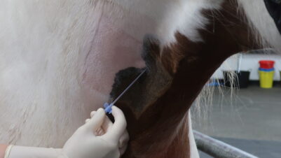
With the development of point-of-care lactate analysers which are relatively inexpensive and readily available, lactate measurements are becoming very widespread in veterinary practice and sampling techniques and handling should be carefully considered in order to obtain consistent and reliable values. It is essential to process blood samples rapidly, as red blood cells are major lactate producers and it has been demonstrated that lactate concentrations increase by 20 percent each hour of storage at room temperature (O’Neill, 2000). Alternatively, samples can be stored on ice or if needed to be sent to an external laboratory they can be stored in tubes containing sodium fluoride which inhibits intracellular glycolytic enzymes (O’Neill, 2000). Sampling sites are also relevant when interpreting plasma lactate values as peripheral venous samples will tend to reflect regional perfusion and arterial samples are the best reflection of the systemic lactate concentration (Hughes et al., 1999). Struggling and prolonged vein occlusion should also be avoided as they can both falsely raise the plasma lactate value (Rand et al., 2002).
In summary
Hyperlactataemia is a common finding in the emergency veterinary patient and should always prompt further investigations to determine the underlying cause and implement appropriate treatment. It is important to realise that a high plasma lactate value is not harmful in itself and should be interpreted as a protective mechanism for the body to preserve cellular energy production. Nevertheless, persistent hyperlactataemia has been associated with higher mortality, so repeated plasma lactate measurements and relative trends are valuable prognostic indicators. Sampling techniques should also be carefully considered when interpreting these results.


