

Feline demodicosis is a disease associated with three Demodex mites. These are Demodex cati, Demodex gatoi and a third unnamed Demodex species.
Demodex cati
This is a long-bodied mite (Figure 1) found in the hair follicles and ears. Disease may be localised or generalised.

Clinical Signs
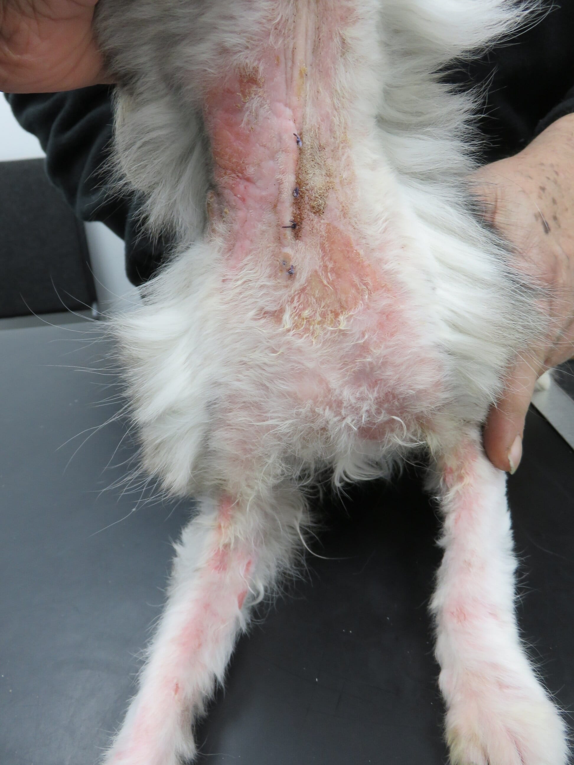
Localised disease involves the eyelids, periocular area, head or neck, and lesions consist of localised alopecia, variable pruritus and erythema. Lesions in generalised disease are multifocal patchy alopecia, with in some cases erythema, scaling, crusts, macules, lichenification and hyper-pigmentation (Figure 2) (Hnilica and Patterson, 2017).
Ceruminous otitis externa alone is also possible. Disease can occur in any age, breed or sex and is not contagious. It may be idiopathic or associated with an underlying immunosuppressive or metabolic disease such as feline immunodeficiency virus (FIV), feline leukaemia virus (FeLV), toxoplasmosis, systemic lupus erythematosus, hyperthyroidism (Figure 2), neoplasia or diabetes mellitus (Hnilica and Patterson, 2017).

Differential diagnosis
In the differential diagnosis, Hnilica and Patterson (2017) include:
- Dermatophytosis
- Cheyletiella
- Otodectes cynotis
- Other causes of otitis externa
Diagnosis
- Skin scrapings. The mite is easier to find than D. gatoi
- Tape strips
- Histopathological examination. Mites may be seen in the stratum corneum or within hair follicles with an associated perifolliculitis and suppurative perivascular dermatitis. Negative findings are not conclusive to rule out of the diagnosis
Treatment
- No specific licence exists for feline demodicosis treatment, therefore informed consent is required
- Lime sulphur 2 percent dips should be applied weekly for four to eight weeks
- Isoxazolines (fluralaner, sarolaner; as with D. gatoi)
- Amitraz dips (0.0125 to 0.025 percent) applied to the whole body weekly to every two weeks (Hnilica and Patterson, 2017). Although reportedly effective, cats are sensitive to the use of amitraz and it is contraindicated in diabetic cats
- Prognosis is variable depending on the underlying cause
Demodex gatoi
This is a short-bodied mite (Figure 4) found in the stratum corneum. Predisposed breeds are Burmese, Maine Coons, Bengals and multi-cat household cats. It is poorly responsive to routine flea treatments, with the exception of the isoxazolines (Matricoti and Maina, 2015).

Clinical signs
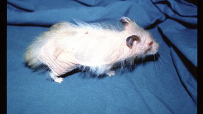
Signs include mild erythema and scaling, which is often generalised. Pruritus is commonly seen and likely due to hypersensitivity to the mite. Signs may be severe and poorly responsive to anti-inflammatory doses of glucocorticoids. Self-induced inflammatory lesions and symmetrical alopecia (Figure 5) may also be seen. Be aware that unlike D. cati, D. gatoi is contagious.
Differential diagnosis
In the differential diagnosis, Hnilica and Patterson (2017) include:
- Dermatophytosis
- Hypersensitivity (Cheyletiella, Otodectes, fleas, food, atopy)
- Psychogenic alopecia

Diagnosis
- Due to excessive licking, the mite may be difficult to find. If there is more than one cat in the household, consider sampling all cats because the mite is easier to find in non-pruritic carrier cats
- Skin scrapings (multiple)
- Tape strips
- Faecal flotation
- Trial therapy is often required
Treatment
- All in-contact cats need treatment
- Fluralaner spot on can be used for cats
- Selamectin and sarolaner spot on (Walker, 2018)
- 2 percent lime sulphur dips. Clinical improvement is often seen within three to four weeks, with a total treatment time of six to eight weeks suggested (Hnilica and Patterson, 2017)
- Prognosis is good
Unnamed Demodex mite
This mite is found in the hair follicles and, in some cases, concurrently with D. cati. It is a distinct species (Ferriera et al., 2015). It has a blunted abdomen and is associated with immunosuppression. The clinical signs and treatment options are similar to those described above for D. cati.


-1628179421-401x226.jpg)
